 Movie
Movie Controller


+ Open data

- Basic information
| Entry | Database: PDB / ID: 3q2w | |||||||||
|---|---|---|---|---|---|---|---|---|---|---|
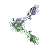
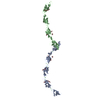
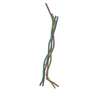
| Title | Crystal structure of mouse N-cadherin ectodomain | |||||||||
 Components Components | Cadherin-2 | |||||||||
 Keywords Keywords | CELL ADHESION / cadherin / calcium binding | |||||||||
| Function / homology |  Function and homology information Function and homology informationmesenchymal cell migration / regulation of oligodendrocyte progenitor proliferation / regulation of postsynaptic density protein 95 clustering / radial glial cell differentiation / neuroligin clustering involved in postsynaptic membrane assembly / positive regulation of synaptic vesicle clustering / Regulation of Insulin-like Growth Factor (IGF) transport and uptake by Insulin-like Growth Factor Binding Proteins (IGFBPs) / Post-translational protein phosphorylation / Adherens junctions interactions / desmosome ...mesenchymal cell migration / regulation of oligodendrocyte progenitor proliferation / regulation of postsynaptic density protein 95 clustering / radial glial cell differentiation / neuroligin clustering involved in postsynaptic membrane assembly / positive regulation of synaptic vesicle clustering / Regulation of Insulin-like Growth Factor (IGF) transport and uptake by Insulin-like Growth Factor Binding Proteins (IGFBPs) / Post-translational protein phosphorylation / Adherens junctions interactions / desmosome / synaptic vesicle clustering / gamma-catenin binding / neural crest cell development / glial cell differentiation / telencephalon development / neuroepithelial cell differentiation / type B pancreatic cell development / cell-cell adhesion mediated by cadherin / neuronal stem cell population maintenance / alpha-catenin binding / fascia adherens / apicolateral plasma membrane / calcium-dependent cell-cell adhesion via plasma membrane cell adhesion molecules / Myogenesis / regulation of Rho protein signal transduction / brain morphogenesis / catenin complex / postsynaptic specialization membrane / cell-cell junction assembly / adherens junction organization / blood vessel morphogenesis / regulation of myelination / heterophilic cell-cell adhesion via plasma membrane cell adhesion molecules / cortical actin cytoskeleton / regulation of axonogenesis / nitric-oxide synthase binding / homophilic cell adhesion via plasma membrane adhesion molecules / plasma membrane raft / intercalated disc / homeostasis of number of cells / presynaptic active zone membrane / regulation of synaptic transmission, glutamatergic / T-tubule / synapse assembly / striated muscle cell differentiation / protein tyrosine kinase binding / protein localization to plasma membrane / adherens junction / brain development / modulation of chemical synaptic transmission / negative regulation of canonical Wnt signaling pathway / cell morphogenesis / sarcolemma / cerebral cortex development / beta-catenin binding / cell-cell adhesion / regulation of protein localization / cell-cell junction / cell migration / apical part of cell / lamellipodium / basolateral plasma membrane / protein phosphatase binding / positive regulation of MAPK cascade / postsynaptic density / cell adhesion / neuron projection / cadherin binding / apical plasma membrane / glutamatergic synapse / synapse / calcium ion binding / protein-containing complex binding / protein kinase binding / enzyme binding / cell surface / protein-containing complex / RNA binding / membrane / identical protein binding / plasma membrane / cytoplasmSimilarity search - Function | |||||||||
| Biological species |  Mus musculus (house mouse) Mus musculus (house mouse) | |||||||||
| Method | X-RAY DIFFRACTION / SYNCHROTRON / MOLECULAR REPLACEMENT / Resolution: 3.2 Å | |||||||||
 Authors Authors | Jin, X. / Shapiro, L. | |||||||||
 Citation Citation | Journal: Structure / Year: 2011 Title: The extracellular architecture of adherens junctions revealed by crystal structures of type I cadherins. Authors: Harrison, O.J. / Jin, X. / Hong, S. / Bahna, F. / Ahlsen, G. / Brasch, J. / Wu, Y. / Vendome, J. / Felsovalyi, K. / Hampton, C.M. / Troyanovsky, R.B. / Ben-Shaul, A. / Frank, J. / ...Authors: Harrison, O.J. / Jin, X. / Hong, S. / Bahna, F. / Ahlsen, G. / Brasch, J. / Wu, Y. / Vendome, J. / Felsovalyi, K. / Hampton, C.M. / Troyanovsky, R.B. / Ben-Shaul, A. / Frank, J. / Troyanovsky, S.M. / Shapiro, L. / Honig, B. | |||||||||
| History |
|
- Structure visualization
| Structure viewer | Molecule: MolmilJmol/JSmol |
|---|
- Downloads & links
-Download
| PDBx/mmCIF format | 3q2w.cif.gz | 124.6 KB | Display | PDBx/mmCIF format |
|---|---|---|---|---|
| PDB format | pdb3q2w.ent.gz | 99 KB | Display | PDB format |
| PDBx/mmJSON format | 3q2w.json.gz | Tree view | PDBx/mmJSON format | |
| Others |  Other downloads Other downloads |
-Validation report
| Arichive directory | https://data.pdbj.org/pub/pdb/validation_reports/q2/3q2wftp://data.pdbj.org/pub/pdb/validation_reports/q2/3q2w | HTTPS FTP |
|---|
-Related structure data


-Links
 PDBj
PDBj


- Assembly
| Deposited unit | 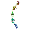
| ||||||||
|---|---|---|---|---|---|---|---|---|---|
| 1 | 
| ||||||||
| Unit cell |
|
-Components
-Protein , 1 types, 1 molecules A
| #1: Protein | / Neural cadherin / N-cadherin Mass: 61529.699 Da / Num. of mol.: 1 / Fragment: UNP residues 160-711 Source method: isolated from a genetically manipulated source Source: (gene. exp.) Mus musculus (house mouse) / Gene: Cdh2 / Cell (production host): HEK293 / Production host:  Homo sapiens (human) / References: UniProt: P15116 Homo sapiens (human) / References: UniProt: P15116 |
|---|
-Sugars , 3 types, 14 molecules 

| #2: Polysaccharide | Source method: isolated from a genetically manipulated source #4: Sugar | ChemComp-MAN / Mannose Type: D-saccharide, alpha linking / Mass: 180.156 Da / Num. of mol.: 9 Type: D-saccharide, alpha linking / Mass: 180.156 Da / Num. of mol.: 9Source method: isolated from a genetically manipulated source Formula: C6H12O6 #5: Sugar | N-Acetylglucosamine Type: D-saccharide, beta linking / Mass: 221.208 Da / Num. of mol.: 3 Type: D-saccharide, beta linking / Mass: 221.208 Da / Num. of mol.: 3Source method: isolated from a genetically manipulated source Formula: C8H15NO6 |
|---|
-Non-polymers , 2 types, 73 molecules 

| #3: Chemical | ChemComp-CA /  Mass: 40.078 Da / Num. of mol.: 12 / Source method: obtained synthetically / Formula: Ca Mass: 40.078 Da / Num. of mol.: 12 / Source method: obtained synthetically / Formula: Ca#6: Water | ChemComp-HOH / | WaterMass: 18.015 Da / Num. of mol.: 61 / Source method: isolated from a natural source / Formula: H2O |
|---|
-Experimental details
-Experiment
| Experiment | Method: X-RAY DIFFRACTION / Number of used crystals: 1 |
|---|
- Sample preparation
| Crystal | Density Matthews: 5.44 Å3/Da / Density % sol: 77.39 % |
|---|---|
| Crystal grow | Temperature: 293 K / Method: vapor diffusion, hanging drop / pH: 8.5 Details: 25% PEG8000, 0.1M Tris, pH 8.5, VAPOR DIFFUSION, HANGING DROP, temperature 293K |
-Data collection
| Diffraction | Mean temperature: 100 K |
|---|---|
| Diffraction source | Source: SYNCHROTRON / Site: NSLS  / Beamline: X4C / Wavelength: 0.9793 Å / Beamline: X4C / Wavelength: 0.9793 Å |
| Detector | Type: MAR CCD 165 mm / Detector: CCD / Date: Jul 31, 2009 |
| Radiation | Monochromator: Si (111) crystal monochromator with vertical focusing mirror Protocol: SINGLE WAVELENGTH / Monochromatic (M) / Laue (L): M / Scattering type: x-ray |
| Radiation wavelength | Wavelength: 0.9793 Å / Relative weight: 1 |
| Reflection | Resolution: 3.2→20 Å / Num. all: 24506 / Num. obs: 21590 / % possible obs: 88.1 % / Observed criterion σ(F): 1 / Observed criterion σ(I): 1 |
- Processing
| Software |
| ||||||||||||||||||||||||||||||||||||||||||||||||||||||||
|---|---|---|---|---|---|---|---|---|---|---|---|---|---|---|---|---|---|---|---|---|---|---|---|---|---|---|---|---|---|---|---|---|---|---|---|---|---|---|---|---|---|---|---|---|---|---|---|---|---|---|---|---|---|---|---|---|---|
| Refinement | Method to determine structure: MOLECULAR REPLACEMENT / Resolution: 3.2→19.947 Å / SU ML: 0.4 / σ(F): 1.34 / Stereochemistry target values: ML
| ||||||||||||||||||||||||||||||||||||||||||||||||||||||||
| Solvent computation | Shrinkage radii: 0.9 Å / VDW probe radii: 1.11 Å / Solvent model: FLAT BULK SOLVENT MODEL / Bsol: 0.796 Å2 / ksol: 0.236 e/Å3 | ||||||||||||||||||||||||||||||||||||||||||||||||||||||||
| Displacement parameters |
| ||||||||||||||||||||||||||||||||||||||||||||||||||||||||
| Refinement step | Cycle: LAST / Resolution: 3.2→19.947 Å
| ||||||||||||||||||||||||||||||||||||||||||||||||||||||||
| Refine LS restraints |
| ||||||||||||||||||||||||||||||||||||||||||||||||||||||||
| LS refinement shell |
|
