 Movie
Movie Controller

[English] 日本語
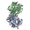
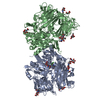
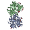
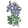
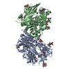
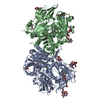
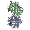
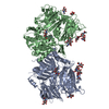
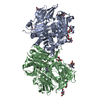
 Yorodumi
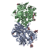
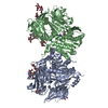
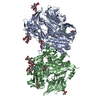
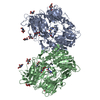
- PDB-3q0t: Crystal structure of human dpp-iv in complex withsa-(+)- methyl2-... -
+ Open data

- Basic information
| Entry | Database: PDB / ID: 3q0t | |||||||||
|---|---|---|---|---|---|---|---|---|---|---|
| Title | Crystal structure of human dpp-iv in complex withsa-(+)- methyl2-(3-(aminomethyl)-4-(2,4-dichlorophenyl)-2-methyl- 7-oxo-5h-pyrrolo[3,4-b]pyridin-6(7h)-yl)acetate | |||||||||
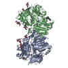
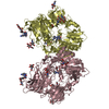
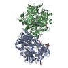
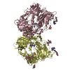
 Components Components | Dipeptidyl peptidase 4 Dipeptidyl peptidase-4 Dipeptidyl peptidase-4 | |||||||||
 Keywords Keywords | hydrolase/hydrolase inhibitor / EXOPEPTIDASE / ALPHA/BETA HYDROLASE FOLD / BETA BARREL / BETA PROPELLER / AMINOPEPTIDASE / GLYCOPROTEIN / MEMBRANE / SERINE PROTEASE / SIGNAL-ANCHOR / TRANSMEMBRANE / hydrolase-hydrolase inhibitor complex | |||||||||
| Function / homology |  Function and homology information Function and homology informationglucagon processing / negative regulation of neutrophil chemotaxis / Synthesis, secretion, and inactivation of Glucose-dependent Insulinotropic Polypeptide (GIP) / regulation of cell-cell adhesion mediated by integrin / negative regulation of extracellular matrix disassembly / psychomotor behavior / dipeptidyl-peptidase IV / intercellular canaliculus / chemorepellent activity / dipeptidyl-peptidase activity ...glucagon processing / negative regulation of neutrophil chemotaxis / Synthesis, secretion, and inactivation of Glucose-dependent Insulinotropic Polypeptide (GIP) / regulation of cell-cell adhesion mediated by integrin / negative regulation of extracellular matrix disassembly / psychomotor behavior / dipeptidyl-peptidase IV / intercellular canaliculus / chemorepellent activity / dipeptidyl-peptidase activity / peptide hormone processing / locomotory exploration behavior / lamellipodium membrane / endocytic vesicle / endothelial cell migration / behavioral fear response / aminopeptidase activity / T cell costimulation / T cell activation / serine-type peptidase activity / Synthesis, secretion, and inactivation of Glucagon-like Peptide-1 (GLP-1) / virus receptor activity / lamellipodium / protease binding / receptor-mediated endocytosis of virus by host cell / membrane fusion / receptor-mediated virion attachment to host cell / response to hypoxia / cell adhesion / symbiont entry into host cell / membrane raft / apical plasma membrane / lysosomal membrane / signaling receptor binding / serine-type endopeptidase activity / focal adhesion / positive regulation of cell population proliferation / cell surface / protein homodimerization activity / proteolysis / extracellular exosome / extracellular region / membrane / identical protein binding / plasma membraneSimilarity search - Function | |||||||||
| Biological species |  Homo sapiens (human) Homo sapiens (human) | |||||||||
| Method | X-RAY DIFFRACTION / SYNCHROTRON / MOLECULAR REPLACEMENT / Resolution: 2.401 Å | |||||||||
 Authors Authors | Klei, H.E. | |||||||||
 Citation Citation | Journal: To be Published Title: Discovery of 7-oxo-pyrrolopyridines as potent and selective inhibitors of dpp4 Authors: Wang, W. / Devasthale, P. / Wang, A. / Egan, D. / Morgan, N. / Cap, M. / Fura, A. / Klei, H.E. / Kish, K. / Sun, L. / Levesque, P. / Zahler, R. / Kirby, M. / Hamann, L.G. | |||||||||
| History |
|
- Structure visualization
| Structure viewer | Molecule: MolmilJmol/JSmol |
|---|
- Downloads & links
-Download
| PDBx/mmCIF format | 3q0t.cif.gz | 316.9 KB | Display | PDBx/mmCIF format |
|---|---|---|---|---|
| PDB format | pdb3q0t.ent.gz | 253.3 KB | Display | PDB format |
| PDBx/mmJSON format | 3q0t.json.gz | Tree view | PDBx/mmJSON format | |
| Others |  Other downloads Other downloads |
-Validation report
| Arichive directory | https://data.pdbj.org/pub/pdb/validation_reports/q0/3q0tftp://data.pdbj.org/pub/pdb/validation_reports/q0/3q0t | HTTPS FTP |
|---|
-Related structure data
| Related structure data | 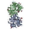 3noxS S: Starting model for refinement |
|---|---|
| Similar structure data |


-Links
 PDBj
PDBj


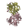
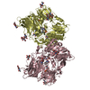
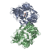
- Assembly
| Deposited unit | 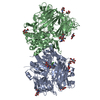
| ||||||||
|---|---|---|---|---|---|---|---|---|---|
| 1 |
| ||||||||
| Unit cell |
|
-Components
| #1: Protein | Dipeptidyl peptidase-4 / ADABP / Adenosine deaminase complexing protein 2 / ADCP-2 / Dipeptidyl peptidase IV / DPP IV / T- ...ADABP / Adenosine deaminase complexing protein 2 / ADCP-2 / Dipeptidyl peptidase IV / DPP IV / T-cell activation antigen CD26 / TP103 / Dipeptidyl peptidase 4 membrane form / Dipeptidyl peptidase IV membrane form / Dipeptidyl peptidase 4 soluble form / Dipeptidyl peptidase IV soluble form Mass: 87450.844 Da / Num. of mol.: 2 Source method: isolated from a genetically manipulated source Source: (gene. exp.) Homo sapiens (human) / Gene: DPP4, ADCP2, CD26 / Plasmid: pPICZalpha / Production host:  Pichia pastoris (fungus) / References: UniProt: P27487, dipeptidyl-peptidase IV Pichia pastoris (fungus) / References: UniProt: P27487, dipeptidyl-peptidase IV#2: Polysaccharide | / Mass: 424.401 Da / Num. of mol.: 2Source method: isolated from a genetically manipulated source #3: Sugar | ChemComp-NAG / N-Acetylglucosamine  Type: D-saccharide, beta linking / Mass: 221.208 Da / Num. of mol.: 10 Type: D-saccharide, beta linking / Mass: 221.208 Da / Num. of mol.: 10Source method: isolated from a genetically manipulated source Formula: C8H15NO6 #4: Chemical |   Mass: 394.252 Da / Num. of mol.: 2 / Source method: obtained synthetically / Formula: C18H17Cl2N3O3 Mass: 394.252 Da / Num. of mol.: 2 / Source method: obtained synthetically / Formula: C18H17Cl2N3O3#5: Water | ChemComp-HOH / | Water Mass: 18.015 Da / Num. of mol.: 431 / Source method: isolated from a natural source / Formula: H2O Mass: 18.015 Da / Num. of mol.: 431 / Source method: isolated from a natural source / Formula: H2O |
|---|
-Experimental details
-Experiment
| Experiment | Method: X-RAY DIFFRACTION / Number of used crystals: 1 |
|---|
- Sample preparation
| Crystal | Density Matthews: 2.67 Å3/Da / Density % sol: 53.94 % |
|---|---|
| Crystal grow | Temperature: 298 K / Method: vapor diffusion, hanging drop / pH: 8.5 Details: CRYSTALS GROWN IN 2 UL EQUIVOLUME MIXTURE OF PROTEIN SOLUTION (0.1 M NACL, 20 MM TRIS-HCL BUFFER PH 7.8, 9.8 MG/ML PROTEIN) AND CRYSTALLIZATION SOLUTION (17% W/V PEG 3350, 15% W/V GLYCEROL, ...Details: CRYSTALS GROWN IN 2 UL EQUIVOLUME MIXTURE OF PROTEIN SOLUTION (0.1 M NACL, 20 MM TRIS-HCL BUFFER PH 7.8, 9.8 MG/ML PROTEIN) AND CRYSTALLIZATION SOLUTION (17% W/V PEG 3350, 15% W/V GLYCEROL, 200 MM MGCL2, 100 MM TRIS-HCL BUFFER PH 8.5). SUFFICIENT. 100 MM LIGAND STOCK SOLUTION (NEAT DMSO) ADDED TO ACHIEVE 1 MM LIGAND CONCENTRATION. SOAKED OVERNIGHT. HARVESTED DIRECTLY AND CRYO-STORED IN LN2. 2. temperature 298K, VAPOR DIFFUSION, HANGING DROP |
-Data collection
| Diffraction source | Source: SYNCHROTRON / Site: NSLS  / Beamline: X29A / Beamline: X29A | |||||||||||||||||||||||||||||||||||||||||||||||||||||||||||||||||||||||||||||
|---|---|---|---|---|---|---|---|---|---|---|---|---|---|---|---|---|---|---|---|---|---|---|---|---|---|---|---|---|---|---|---|---|---|---|---|---|---|---|---|---|---|---|---|---|---|---|---|---|---|---|---|---|---|---|---|---|---|---|---|---|---|---|---|---|---|---|---|---|---|---|---|---|---|---|---|---|---|---|
| Radiation | Protocol: SINGLE WAVELENGTH / Monochromatic (M) / Laue (L): M / Scattering type: x-ray | |||||||||||||||||||||||||||||||||||||||||||||||||||||||||||||||||||||||||||||
| Radiation wavelength | Relative weight: 1 | |||||||||||||||||||||||||||||||||||||||||||||||||||||||||||||||||||||||||||||
| Reflection | Resolution: 2.4→50 Å / Num. obs: 71882 / % possible obs: 96.1 % / Redundancy: 7.3 % / Rmerge(I) obs: 0.068 / Χ2: 0.938 / Net I/σ(I): 10.6 | |||||||||||||||||||||||||||||||||||||||||||||||||||||||||||||||||||||||||||||
| Reflection shell |
|
- Processing
| Software |
| ||||||||||||||||||||||||||||||||||||||||||||||||||||||||||||||||||||||||||||||||||||||||||||||||||||||||||||||||||||||||||||||||||||||||||||||||||||||||||
|---|---|---|---|---|---|---|---|---|---|---|---|---|---|---|---|---|---|---|---|---|---|---|---|---|---|---|---|---|---|---|---|---|---|---|---|---|---|---|---|---|---|---|---|---|---|---|---|---|---|---|---|---|---|---|---|---|---|---|---|---|---|---|---|---|---|---|---|---|---|---|---|---|---|---|---|---|---|---|---|---|---|---|---|---|---|---|---|---|---|---|---|---|---|---|---|---|---|---|---|---|---|---|---|---|---|---|---|---|---|---|---|---|---|---|---|---|---|---|---|---|---|---|---|---|---|---|---|---|---|---|---|---|---|---|---|---|---|---|---|---|---|---|---|---|---|---|---|---|---|---|---|---|---|---|---|
| Refinement | Method to determine structure: MOLECULAR REPLACEMENT Starting model: 3NOX Resolution: 2.401→47.952 Å / Occupancy max: 1 / Occupancy min: 1 / SU ML: 0.34 / σ(F): 1.44 / Stereochemistry target values: ML
| ||||||||||||||||||||||||||||||||||||||||||||||||||||||||||||||||||||||||||||||||||||||||||||||||||||||||||||||||||||||||||||||||||||||||||||||||||||||||||
| Solvent computation | Shrinkage radii: 0.9 Å / VDW probe radii: 1.11 Å / Solvent model: FLAT BULK SOLVENT MODEL / Bsol: 42.697 Å2 / ksol: 0.324 e/Å3 | ||||||||||||||||||||||||||||||||||||||||||||||||||||||||||||||||||||||||||||||||||||||||||||||||||||||||||||||||||||||||||||||||||||||||||||||||||||||||||
| Displacement parameters | Biso max: 94.99 Å2 / Biso mean: 44.3494 Å2 / Biso min: 19.11 Å2
| ||||||||||||||||||||||||||||||||||||||||||||||||||||||||||||||||||||||||||||||||||||||||||||||||||||||||||||||||||||||||||||||||||||||||||||||||||||||||||
| Refinement step | Cycle: LAST / Resolution: 2.401→47.952 Å
| ||||||||||||||||||||||||||||||||||||||||||||||||||||||||||||||||||||||||||||||||||||||||||||||||||||||||||||||||||||||||||||||||||||||||||||||||||||||||||
| Refine LS restraints |
| ||||||||||||||||||||||||||||||||||||||||||||||||||||||||||||||||||||||||||||||||||||||||||||||||||||||||||||||||||||||||||||||||||||||||||||||||||||||||||
| LS refinement shell | Refine-ID: X-RAY DIFFRACTION / Total num. of bins used: 21
|
