 Movie
Movie Controller

[English] 日本語
 Yorodumi
- PDB-3q05: An induced fit mechanism regulates p53 DNA binding kinetics to co... -
+ Open data

- Basic information
| Entry | Database: PDB / ID: 3q05 | ||||||
|---|---|---|---|---|---|---|---|
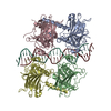
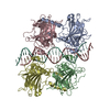
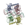
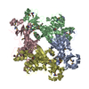
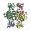
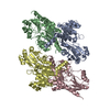
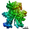
| Title | An induced fit mechanism regulates p53 DNA binding kinetics to confer sequence specificity | ||||||
 Components Components |
| ||||||
 Keywords Keywords |  ANTITUMOR PROTEIN/DNA / beta sandwich / multidomain / oligomerization / TP53 / p53 / tumor suppressor / tetramer / DNA binding / ANTITUMOR PROTEIN-DNA complex ANTITUMOR PROTEIN/DNA / beta sandwich / multidomain / oligomerization / TP53 / p53 / tumor suppressor / tetramer / DNA binding / ANTITUMOR PROTEIN-DNA complex | ||||||
| Function / homology |  Function and homology information Function and homology informationLoss of function of TP53 in cancer due to loss of tetramerization ability / Regulation of TP53 Expression / signal transduction by p53 class mediator / negative regulation of G1 to G0 transition / negative regulation of glucose catabolic process to lactate via pyruvate / Transcriptional activation of cell cycle inhibitor p21 / regulation of intrinsic apoptotic signaling pathway by p53 class mediator / Activation of NOXA and translocation to mitochondria / negative regulation of pentose-phosphate shunt / ATP-dependent DNA/DNA annealing activity ...Loss of function of TP53 in cancer due to loss of tetramerization ability / Regulation of TP53 Expression / signal transduction by p53 class mediator / negative regulation of G1 to G0 transition / negative regulation of glucose catabolic process to lactate via pyruvate / Transcriptional activation of cell cycle inhibitor p21 / regulation of intrinsic apoptotic signaling pathway by p53 class mediator / Activation of NOXA and translocation to mitochondria / negative regulation of pentose-phosphate shunt / ATP-dependent DNA/DNA annealing activity / negative regulation of helicase activity / regulation of cell cycle G2/M phase transition / intrinsic apoptotic signaling pathway in response to hypoxia / regulation of fibroblast apoptotic process / oxidative stress-induced premature senescence / oligodendrocyte apoptotic process / negative regulation of miRNA processing / positive regulation of thymocyte apoptotic process / glucose catabolic process to lactate via pyruvate / regulation of tissue remodeling / positive regulation of mitochondrial membrane permeability / negative regulation of mitophagy / positive regulation of programmed necrotic cell death / mRNA transcription / bone marrow development / circadian behavior / histone deacetylase regulator activity / regulation of mitochondrial membrane permeability involved in apoptotic process / RUNX3 regulates CDKN1A transcription / germ cell nucleus / regulation of DNA damage response, signal transduction by p53 class mediator / TP53 regulates transcription of additional cell cycle genes whose exact role in the p53 pathway remain uncertain / TP53 Regulates Transcription of Death Receptors and Ligands / Activation of PUMA and translocation to mitochondria / DNA damage response, signal transduction by p53 class mediator resulting in transcription of p21 class mediator / negative regulation of glial cell proliferation / Formation of Senescence-Associated Heterochromatin Foci (SAHF) / negative regulation of neuroblast proliferation / Regulation of TP53 Activity through Association with Co-factors / mitochondrial DNA repair / T cell lineage commitment / positive regulation of execution phase of apoptosis / negative regulation of DNA replication / ER overload response / B cell lineage commitment / thymocyte apoptotic process / TP53 regulates transcription of several additional cell death genes whose specific roles in p53-dependent apoptosis remain uncertain / positive regulation of cardiac muscle cell apoptotic process / TP53 Regulates Transcription of Caspase Activators and Caspases / T cell proliferation involved in immune response / cardiac septum morphogenesis / entrainment of circadian clock by photoperiod / PI5P Regulates TP53 Acetylation / Association of TriC/CCT with target proteins during biosynthesis / Zygotic genome activation (ZGA) / necroptotic process / rRNA transcription / positive regulation of release of cytochrome c from mitochondria / TP53 Regulates Transcription of Genes Involved in Cytochrome C Release / TFIID-class transcription factor complex binding / mitophagy / SUMOylation of transcription factors / negative regulation of telomere maintenance via telomerase / general transcription initiation factor binding / intrinsic apoptotic signaling pathway by p53 class mediator / Transcriptional Regulation by VENTX / response to X-ray / DNA damage response, signal transduction by p53 class mediator / replicative senescence / chromosome organization / neuroblast proliferation / intrinsic apoptotic signaling pathway in response to endoplasmic reticulum stress / cellular response to UV-C / : / hematopoietic stem cell differentiation / negative regulation of reactive oxygen species metabolic process / intrinsic apoptotic signaling pathway in response to DNA damage by p53 class mediator / glial cell proliferation / embryonic organ development / positive regulation of RNA polymerase II transcription preinitiation complex assembly / Pyroptosis / cis-regulatory region sequence-specific DNA binding / hematopoietic progenitor cell differentiation / cellular response to glucose starvation / cellular response to actinomycin D / TP53 Regulates Transcription of Genes Involved in G1 Cell Cycle Arrest / somitogenesis / type II interferon-mediated signaling pathway / positive regulation of intrinsic apoptotic signaling pathway / negative regulation of stem cell proliferation / core promoter sequence-specific DNA binding / gastrulation / negative regulation of fibroblast proliferation / MDM2/MDM4 family protein binding / cardiac muscle cell apoptotic process / transcription initiation-coupled chromatin remodeling / 14-3-3 protein binding / mitotic G1 DNA damage checkpoint signaling / Regulation of TP53 Activity through Acetylation / response to salt stressSimilarity search - Function | ||||||
| Biological species |  Homo sapiens (human) Homo sapiens (human) | ||||||
| Method | X-RAY DIFFRACTION / SYNCHROTRON / MOLECULAR REPLACEMENT / Resolution: 2.4 Å | ||||||
 Authors Authors | Petty, T.J. / Halazonetis, T.D. | ||||||
 Citation Citation | Journal: Embo J. / Year: 2011 Title: An induced fit mechanism regulates p53 DNA binding kinetics to confer sequence specificity. Authors: Petty, T.J. / Emamzadah, S. / Costantino, L. / Petkova, I. / Stavridi, E.S. / Saven, J.G. / Vauthey, E. / Halazonetis, T.D. | ||||||
| History |
|
- Structure visualization
| Structure viewer | Molecule: MolmilJmol/JSmol |
|---|
- Downloads & links
-Download
| PDBx/mmCIF format | 3q05.cif.gz | 226.3 KB | Display | PDBx/mmCIF format |
|---|---|---|---|---|
| PDB format | pdb3q05.ent.gz | 177.1 KB | Display | PDB format |
| PDBx/mmJSON format | 3q05.json.gz | Tree view | PDBx/mmJSON format | |
| Others |  Other downloads Other downloads |
-Validation report
| Arichive directory | https://data.pdbj.org/pub/pdb/validation_reports/q0/3q05ftp://data.pdbj.org/pub/pdb/validation_reports/q0/3q05 | HTTPS FTP |
|---|
-Related structure data
| Related structure data | 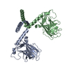 3q01C  3q06C  1c26S 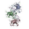 1tupS C: citing same article ( S: Starting model for refinement |
|---|---|
| Similar structure data |

-Links
 PDBj
PDBj


- Assembly
| Deposited unit | 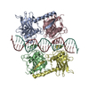
| ||||||||
|---|---|---|---|---|---|---|---|---|---|
| 1 |
| ||||||||
| Unit cell |
|
-Components
| #1: Protein | P53 / Antigen NY-CO-13 / Phosphoprotein p53 / Tumor suppressor p53 Mass: 26618.098 Da / Num. of mol.: 4 Fragment: p53 DNA-binding (Res 94-291) and Oligomerization (Res 321-356) domains Mutation: C135V, C141V, W146Y, C182S, V203A, R209P, C229Y, H233Y, Y234F, N235K, Y236F, T253V, N268D, P322A, L323M, M340Q, L344R, G356T Source method: isolated from a genetically manipulated source Source: (gene. exp.) Homo sapiens (human) / Gene: TP53, p53 / Plasmid: pT5T / Production host:  Escherichia coli (E. coli) / Strain (production host): BL21(DE3) / References: UniProt: P04637 Escherichia coli (E. coli) / Strain (production host): BL21(DE3) / References: UniProt: P04637#2: DNA chain | | Mass: 7948.129 Da / Num. of mol.: 1 / Source method: obtained synthetically / Details: Consensus human TP53 binding sequence sense strand #3: DNA chain | | Mass: 8028.178 Da / Num. of mol.: 1 / Source method: obtained synthetically Details: Consensus human TP53 binding sequence anti-sense strand #4: Chemical | ChemComp-ZN /   Mass: 65.409 Da / Num. of mol.: 4 / Source method: obtained synthetically / Formula: Zn Mass: 65.409 Da / Num. of mol.: 4 / Source method: obtained synthetically / Formula: Zn#5: Water | ChemComp-HOH / | Water Mass: 18.015 Da / Num. of mol.: 161 / Source method: isolated from a natural source / Formula: H2O Mass: 18.015 Da / Num. of mol.: 161 / Source method: isolated from a natural source / Formula: H2O |
|---|
-Experimental details
-Experiment
| Experiment | Method: X-RAY DIFFRACTION / Number of used crystals: 1 |
|---|
- Sample preparation
| Crystal | Density Matthews: 3.15 Å3/Da / Density % sol: 60.96 % |
|---|---|
| Crystal grow | Temperature: 277 K / Method: vapor diffusion, hanging drop / pH: 7 Details: 0.15M DL-Malic Acid [pH 7.0], 20% Polyethylene Glycol 3350, VAPOR DIFFUSION, HANGING DROP, temperature 277K |
-Data collection
| Diffraction | Mean temperature: 85 K |
|---|---|
| Diffraction source | Source: SYNCHROTRON / Site: ESRF  / Beamline: ID23-2 / Wavelength: 0.8726 Å / Beamline: ID23-2 / Wavelength: 0.8726 Å |
| Detector | Type: MARMOSAIC 225 mm CCD / Detector: CCD / Date: Feb 13, 2009 |
| Radiation | Monochromator: Pt coated Si mirror / Protocol: SINGLE WAVELENGTH / Monochromatic (M) / Laue (L): M / Scattering type: x-ray |
| Radiation wavelength | Wavelength: 0.8726 Å / Relative weight: 1 |
| Reflection | Resolution: 2.4→41.07 Å / Num. all: 59945 / Num. obs: 59874 / % possible obs: 98.6 % / Observed criterion σ(F): 0 / Observed criterion σ(I): 0 / Rsym value: 0.117 / Net I/σ(I): 10.9 |
| Reflection shell | Resolution: 2.4→2.53 Å / Mean I/σ(I) obs: 2.5 / Num. unique all: 8752 / Rsym value: 0.442 / % possible all: 98.6 |
- Processing
| Software |
| |||||||||||||||||||||||||
|---|---|---|---|---|---|---|---|---|---|---|---|---|---|---|---|---|---|---|---|---|---|---|---|---|---|---|
| Refinement | Method to determine structure: MOLECULAR REPLACEMENT Starting model: PDB entries 1TUP, 1C26 Resolution: 2.4→41.07 Å / Isotropic thermal model: Overall / Cross valid method: THROUGHOUT / σ(F): 0.5 / Stereochemistry target values: Engh & Huber
| |||||||||||||||||||||||||
| Refinement step | Cycle: LAST / Resolution: 2.4→41.07 Å
| |||||||||||||||||||||||||
| Refine LS restraints |
|
