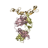
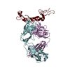
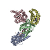
Mass: 74353.219 Da / Num. of mol.: 1 / Fragment: MyTH4-FERM-SH3 region Source method: isolated from a genetically manipulated source Source: (gene. exp.) Mus musculus (house mouse) / Gene: Myo7a / Production host: Escherichia coli (E. coli) / Strain (production host): BL21 / References: UniProt: Q5MJ57, UniProt: P97479*PLUS
#2: Protein
Ushersyndrometype-1Gprotein / Scaffold protein containing ankyrin repeats and SAM domain
Mass: 10215.873 Da / Num. of mol.: 1 / Fragment: Central region of Sans, the CEN1 motif Source method: isolated from a genetically manipulated source Source: (gene. exp.) Homo sapiens (human) / Gene: USH1G, SANS / Production host: Escherichia coli (E. coli) / Strain (production host): BL21 / References: UniProt: Q495M9
Resolution: 2.801→2.874 Å / Redundancy: 9.3 % / Rmerge(I) obs: 0.06 / Mean I/σ(I) obs: 31 / Num. unique all: 34633 / % possible all: 99.69
-
Processing
Software
Name
Version
Classification
HKL-2000
datacollection
SHARP
phasing
REFMAC
5.5.0072
refinement
HKL-2000
datareduction
HKL-2000
datascaling
Refinement
Method to determine structure: SAD / Resolution: 2.8→50 Å / Cor.coef. Fo:Fc: 0.932 / Cor.coef. Fo:Fc free: 0.915 / SU B: 11.315 / SU ML: 0.222 / Cross valid method: THROUGHOUT / σ(F): 3.7 / σ(I): 3.7 / ESU R Free: 0.289 / Stereochemistry target values: MAXIMUM LIKELIHOOD / Details: HYDROGENS HAVE BEEN ADDED IN THE RIDING POSITIONS
Rfactor
Num. reflection
% reflection
Selection details
Rfree
0.25903
1740
5 %
RANDOM
Rwork
0.22764
-
-
-
all
0.22922
34633
-
-
obs
0.22922
32761
99.57 %
-
Solvent computation
Ion probe radii: 0.8 Å / Shrinkage radii: 0.8 Å / VDW probe radii: 1.2 Å / Solvent model: BABINET MODEL WITH MASK
Displacement parameters
Biso mean: 67.928 Å2
Baniso -1
Baniso -2
Baniso -3
1-
0.17 Å2
0.08 Å2
0 Å2
2-
-
0.17 Å2
0 Å2
3-
-
-
-0.25 Å2
Refinement step
Cycle: LAST / Resolution: 2.8→50 Å
Protein
Nucleic acid
Ligand
Solvent
Total
Num. atoms
4888
0
64
38
4990
Refine LS restraints
Refine-ID
Type
Dev ideal
Dev ideal target
Number
X-RAY DIFFRACTION
r_bond_refined_d
0.007
0.022
5066
X-RAY DIFFRACTION
r_angle_refined_deg
0.98
1.972
6852
X-RAY DIFFRACTION
r_dihedral_angle_1_deg
4.92
5
610
X-RAY DIFFRACTION
r_dihedral_angle_2_deg
34.239
23.465
228
X-RAY DIFFRACTION
r_dihedral_angle_3_deg
16.836
15
855
X-RAY DIFFRACTION
r_dihedral_angle_4_deg
14.926
15
35
X-RAY DIFFRACTION
r_chiral_restr
0.069
0.2
748
X-RAY DIFFRACTION
r_gen_planes_refined
0.004
0.021
3799
X-RAY DIFFRACTION
r_mcbond_it
1.402
2
3062
X-RAY DIFFRACTION
r_mcangle_it
2.645
3
4950
X-RAY DIFFRACTION
r_scbond_it
3.38
4
2004
X-RAY DIFFRACTION
r_scangle_it
5.585
6
1901
LS refinement shell
Resolution: 2.801→2.874 Å / Total num. of bins used: 20
Rfactor
Num. reflection
% reflection
Rfree
0.39
134
-
Rwork
0.352
2418
-
obs
-
-
99.69 %
+
About Yorodumi
-
News
-
Feb 9, 2022. New format data for meta-information of EMDB entries
New format data for meta-information of EMDB entries
Version 3 of the EMDB header file is now the official format.
The previous official version 1.9 will be removed from the archive.
In the structure databanks used in Yorodumi, some data are registered as the other names, "COVID-19 virus" and "2019-nCoV". Here are the details of the virus and the list of structure data.
Jan 31, 2019. EMDB accession codes are about to change! (news from PDBe EMDB page)
EMDB accession codes are about to change! (news from PDBe EMDB page)
The allocation of 4 digits for EMDB accession codes will soon come to an end. Whilst these codes will remain in use, new EMDB accession codes will include an additional digit and will expand incrementally as the available range of codes is exhausted. The current 4-digit format prefixed with “EMD-” (i.e. EMD-XXXX) will advance to a 5-digit format (i.e. EMD-XXXXX), and so on. It is currently estimated that the 4-digit codes will be depleted around Spring 2019, at which point the 5-digit format will come into force.
The EM Navigator/Yorodumi systems omit the EMD- prefix.
Related info.:Q: What is EMD? / ID/Accession-code notation in Yorodumi/EM Navigator
Yorodumi is a browser for structure data from EMDB, PDB, SASBDB, etc.
This page is also the successor to EM Navigator detail page, and also detail information page/front-end page for Omokage search.
The word "yorodu" (or yorozu) is an old Japanese word meaning "ten thousand". "mi" (miru) is to see.
Related info.:EMDB / PDB / SASBDB / Comparison of 3 databanks / Yorodumi Search / Aug 31, 2016. New EM Navigator & Yorodumi / Yorodumi Papers / Jmol/JSmol / Function and homology information / Changes in new EM Navigator and Yorodumi
 Movie
Movie Controller

 Yorodumi
 Open data
 Basic information
 Components
 Keywords
 MOTOR PROTEIN/PROTEIN TRANSPORT /
  Function and homology information

 Authors
 Citation
 Structure visualization
 Downloads & links
 Other downloads


 PDBj
PDBj


 Assembly


 Mass: 92.094 Da / Num. of mol.: 9 / Source method: obtained synthetically / Formula: C3H8O3
 Mass: 94.971 Da / Num. of mol.: 2 / Source method: obtained synthetically / Formula: PO4
 Mass: 18.015 Da / Num. of mol.: 38 / Source method: isolated from a natural source / Formula: H2O
 Sample preparation
 / Beamline: BL17U / Wavelength: 1.5418 Å
 Processing