 Movie
Movie Controller

[English] 日本語
 Yorodumi
- PDB-3pr6: Crystal structure analysis of yeast TRAPP associate protein Tca17 -
+ Open data

- Basic information
| Entry | Database: PDB / ID: 3pr6 | ||||||
|---|---|---|---|---|---|---|---|
| Title | Crystal structure analysis of yeast TRAPP associate protein Tca17 | ||||||
 Components Components | TRAPP-associated protein TCA17 | ||||||
 Keywords Keywords |  TRANSPORT PROTEIN / longin fold / vesicle tethering regulation / TRAPP complex / trans-Golgi network TRANSPORT PROTEIN / longin fold / vesicle tethering regulation / TRAPP complex / trans-Golgi network | ||||||
| Function / homology |  Function and homology information Function and homology informationTRAPPII protein complex / TRAPP complex / early endosome to Golgi transport / intra-Golgi vesicle-mediated transport / retrograde transport, endosome to Golgi / endoplasmic reticulum to Golgi vesicle-mediated transport / protein-containing complex assembly / Golgi apparatus / nucleus / cytosol / cytoplasmSimilarity search - Function | ||||||
| Biological species |  Saccharomyces cerevisiae (brewer's yeast) Saccharomyces cerevisiae (brewer's yeast) | ||||||
| Method | X-RAY DIFFRACTION / SYNCHROTRON / MAD / Resolution: 1.8 Å | ||||||
 Authors Authors | Wang, C. / Gohlke, U. / Heinemann, U. | ||||||
 Citation Citation | Journal: Febs J. / Year: 2014 Title: Crystal structure of the yeast TRAPP-associated protein Tca17. Authors: Wang, C. / Gohlke, U. / Roske, Y. / Heinemann, U. | ||||||
| History |
|
- Structure visualization
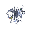
| Structure viewer | Molecule: MolmilJmol/JSmol |
|---|
- Downloads & links
-Download
| PDBx/mmCIF format | 3pr6.cif.gz | 73 KB | Display | PDBx/mmCIF format |
|---|---|---|---|---|
| PDB format | pdb3pr6.ent.gz | 57.3 KB | Display | PDB format |
| PDBx/mmJSON format | 3pr6.json.gz | Tree view | PDBx/mmJSON format | |
| Others |  Other downloads Other downloads |
-Validation report
| Arichive directory | https://data.pdbj.org/pub/pdb/validation_reports/pr/3pr6ftp://data.pdbj.org/pub/pdb/validation_reports/pr/3pr6 | HTTPS FTP |
|---|
-Related structure data
| Similar structure data |
|---|


-Links
 PDBj
PDBj
- Assembly
| Deposited unit | 
| ||||||||
|---|---|---|---|---|---|---|---|---|---|
| 1 |
| ||||||||
| 2 | 
| ||||||||
| Unit cell |
|
-Components
| #1: Protein | Mass: 18352.621 Da / Num. of mol.: 1 Source method: isolated from a genetically manipulated source Source: (gene. exp.) Saccharomyces cerevisiae (brewer's yeast)Gene: SYGP-ORF36, TCA17, YEL048C / Plasmid: pGex6p-1 / Production host:  Escherichia coli (E. coli) / Strain (production host): BL21(DE3)-T1R / References: UniProt: P32613 Escherichia coli (E. coli) / Strain (production host): BL21(DE3)-T1R / References: UniProt: P32613 |
|---|---|
| #2: Chemical | ChemComp-CL / Chloride  Mass: 35.453 Da / Num. of mol.: 1 / Source method: obtained synthetically / Formula: Cl Mass: 35.453 Da / Num. of mol.: 1 / Source method: obtained synthetically / Formula: Cl |
| #3: Chemical | ChemComp-GOL / Glycerol  Mass: 92.094 Da / Num. of mol.: 1 / Source method: obtained synthetically / Formula: C3H8O3 Mass: 92.094 Da / Num. of mol.: 1 / Source method: obtained synthetically / Formula: C3H8O3 |
| #4: Water | ChemComp-HOH / Water Mass: 18.015 Da / Num. of mol.: 133 / Source method: isolated from a natural source / Formula: H2O Mass: 18.015 Da / Num. of mol.: 133 / Source method: isolated from a natural source / Formula: H2O |
-Experimental details
-Experiment
| Experiment | Method: X-RAY DIFFRACTION / Number of used crystals: 2 |
|---|
- Sample preparation
| Crystal | Density Matthews: 2.47 Å3/Da / Density % sol: 50.14 % |
|---|---|
| Crystal grow | Temperature: 277 K / Method: vapor diffusion, sitting drop / pH: 8.5 Details: 25% PEG3350, 0.2 M magnesium chloride, 0.1 M Tris-HCl, pH 8.5, VAPOR DIFFUSION, SITTING DROP, temperature 277K |
-Data collection
| Diffraction | Mean temperature: 100 K | |||||||||||||||||||||||||||||||||||||||||||||||||||||||||||||||||||||||||||||||||||||||||||||||||||||||||||||||||||||||||||||||||||||||||||||||||||
|---|---|---|---|---|---|---|---|---|---|---|---|---|---|---|---|---|---|---|---|---|---|---|---|---|---|---|---|---|---|---|---|---|---|---|---|---|---|---|---|---|---|---|---|---|---|---|---|---|---|---|---|---|---|---|---|---|---|---|---|---|---|---|---|---|---|---|---|---|---|---|---|---|---|---|---|---|---|---|---|---|---|---|---|---|---|---|---|---|---|---|---|---|---|---|---|---|---|---|---|---|---|---|---|---|---|---|---|---|---|---|---|---|---|---|---|---|---|---|---|---|---|---|---|---|---|---|---|---|---|---|---|---|---|---|---|---|---|---|---|---|---|---|---|---|---|---|---|---|
| Diffraction source | Source: SYNCHROTRON / Site: BESSY  / Beamline: 14.2 / Wavelength: 0.9184 / Wavelength: 0.9796,0.9798,0.9720 / Beamline: 14.2 / Wavelength: 0.9184 / Wavelength: 0.9796,0.9798,0.9720 | |||||||||||||||||||||||||||||||||||||||||||||||||||||||||||||||||||||||||||||||||||||||||||||||||||||||||||||||||||||||||||||||||||||||||||||||||||
| Detector | Type: RAYONIX MX-225 / Detector: CCD / Date: May 12, 2010 | |||||||||||||||||||||||||||||||||||||||||||||||||||||||||||||||||||||||||||||||||||||||||||||||||||||||||||||||||||||||||||||||||||||||||||||||||||
| Radiation | Monochromator: Si-111 / Protocol: MAD / Monochromatic (M) / Laue (L): M / Scattering type: x-ray | |||||||||||||||||||||||||||||||||||||||||||||||||||||||||||||||||||||||||||||||||||||||||||||||||||||||||||||||||||||||||||||||||||||||||||||||||||
| Radiation wavelength |
| |||||||||||||||||||||||||||||||||||||||||||||||||||||||||||||||||||||||||||||||||||||||||||||||||||||||||||||||||||||||||||||||||||||||||||||||||||
| Reflection | Resolution: 1.8→29.25 Å / Num. obs: 16453 / % possible obs: 99.4 % / Observed criterion σ(I): -3 / Biso Wilson estimate: 35.308 Å2 / Rmerge(I) obs: 0.039 / Net I/σ(I): 17.49 | |||||||||||||||||||||||||||||||||||||||||||||||||||||||||||||||||||||||||||||||||||||||||||||||||||||||||||||||||||||||||||||||||||||||||||||||||||
| Reflection shell | Diffraction-ID: 1
|
-Phasing
| Phasing | Method: MAD |
|---|
- Processing
| Software |
| ||||||||||||||||||||||||||||||||||||||||||||||||||||||||||||||||||||||||||||||||||||||||||||||||||||
|---|---|---|---|---|---|---|---|---|---|---|---|---|---|---|---|---|---|---|---|---|---|---|---|---|---|---|---|---|---|---|---|---|---|---|---|---|---|---|---|---|---|---|---|---|---|---|---|---|---|---|---|---|---|---|---|---|---|---|---|---|---|---|---|---|---|---|---|---|---|---|---|---|---|---|---|---|---|---|---|---|---|---|---|---|---|---|---|---|---|---|---|---|---|---|---|---|---|---|---|---|---|
| Refinement | Method to determine structure: MAD / Resolution: 1.8→29.25 Å / Cor.coef. Fo:Fc: 0.96 / Cor.coef. Fo:Fc free: 0.944 / WRfactor Rfree: 0.2274 / WRfactor Rwork: 0.1874 / Occupancy max: 1 / Occupancy min: 0.3 / FOM work R set: 0.8785 / SU B: 5.206 / SU ML: 0.069 / SU R Cruickshank DPI: 0.0257 / SU Rfree: 0.025 / Cross valid method: THROUGHOUT / σ(F): 0 / ESU R Free: 0.025 / Stereochemistry target values: MAXIMUM LIKELIHOOD / Details: HYDROGENS HAVE BEEN ADDED IN THE RIDING POSITIONS
| ||||||||||||||||||||||||||||||||||||||||||||||||||||||||||||||||||||||||||||||||||||||||||||||||||||
| Solvent computation | Ion probe radii: 0.8 Å / Shrinkage radii: 0.8 Å / VDW probe radii: 1.4 Å / Solvent model: BABINET MODEL WITH MASK | ||||||||||||||||||||||||||||||||||||||||||||||||||||||||||||||||||||||||||||||||||||||||||||||||||||
| Displacement parameters | Biso max: 73.75 Å2 / Biso mean: 31.76 Å2 / Biso min: 11.17 Å2
| ||||||||||||||||||||||||||||||||||||||||||||||||||||||||||||||||||||||||||||||||||||||||||||||||||||
| Refinement step | Cycle: LAST / Resolution: 1.8→29.25 Å
| ||||||||||||||||||||||||||||||||||||||||||||||||||||||||||||||||||||||||||||||||||||||||||||||||||||
| Refine LS restraints |
| ||||||||||||||||||||||||||||||||||||||||||||||||||||||||||||||||||||||||||||||||||||||||||||||||||||
| LS refinement shell | Resolution: 1.799→1.846 Å / Total num. of bins used: 20
| ||||||||||||||||||||||||||||||||||||||||||||||||||||||||||||||||||||||||||||||||||||||||||||||||||||
| Refinement TLS params. | Method: refined / Refine-ID: X-RAY DIFFRACTION
| ||||||||||||||||||||||||||||||||||||||||||||||||||||||||||||||||||||||||||||||||||||||||||||||||||||
| Refinement TLS group |
|
