 Movie
Movie Controller

[English] 日本語
 Yorodumi
- PDB-3ppg: Crystal structure of the Candida albicans methionine synthase by ... -
+ Open data

- Basic information
| Entry | Database: PDB / ID: 3ppg | ||||||
|---|---|---|---|---|---|---|---|
| Title | Crystal structure of the Candida albicans methionine synthase by surface entropy reduction, alanine variant with zinc | ||||||
 Components Components | 5-methyltetrahydropteroyltriglutamate--homocysteine methyltransferase | ||||||
 Keywords Keywords |  TRANSFERASE / Cobalamin-Independent / Surface Entropy Reduction / Methyltransferase / Metalloproteinase TRANSFERASE / Cobalamin-Independent / Surface Entropy Reduction / Methyltransferase / Metalloproteinase | ||||||
| Function / homology |  Function and homology information Function and homology informationL-methionine biosynthetic process from homoserine via O-acetyl-L-homoserine and cystathionine / : / 5-methyltetrahydropteroyltriglutamate-homocysteine S-methyltransferase / 5-methyltetrahydropteroyltriglutamate-homocysteine S-methyltransferase activity / fungal biofilm matrix / hyphal cell wall / adenine biosynthetic process / methionine synthase activity / methionine metabolic process / 'de novo' L-methionine biosynthetic process ...L-methionine biosynthetic process from homoserine via O-acetyl-L-homoserine and cystathionine / : / 5-methyltetrahydropteroyltriglutamate-homocysteine S-methyltransferase / 5-methyltetrahydropteroyltriglutamate-homocysteine S-methyltransferase activity / fungal biofilm matrix / hyphal cell wall / adenine biosynthetic process / methionine synthase activity / methionine metabolic process / 'de novo' L-methionine biosynthetic process / fungal-type cell wall / methionine biosynthetic process / cellular response to heat / methylation / cell surface / zinc ion binding / extracellular region / nucleus / cytosolSimilarity search - Function | ||||||
| Biological species |  Candida albicans (yeast) Candida albicans (yeast) | ||||||
| Method | X-RAY DIFFRACTION / SYNCHROTRON / MOLECULAR REPLACEMENT / Resolution: 1.98 Å | ||||||
 Authors Authors | Ubhi, D. / Kavanagh, K. / Monzingo, A.F. / Robertus, J.D. | ||||||
 Citation Citation | Journal: Arch.Biochem.Biophys. / Year: 2011 Title: Structure of Candida albicans methionine synthase determined by employing surface residue mutagenesis. Authors: Ubhi, D. / Kavanagh, K.L. / Monzingo, A.F. / Robertus, J.D. | ||||||
| History |
|
- Structure visualization
| Structure viewer | Molecule: MolmilJmol/JSmol |
|---|
- Downloads & links
-Download
| PDBx/mmCIF format | 3ppg.cif.gz | 304.7 KB | Display | PDBx/mmCIF format |
|---|---|---|---|---|
| PDB format | pdb3ppg.ent.gz | 241.4 KB | Display | PDB format |
| PDBx/mmJSON format | 3ppg.json.gz | Tree view | PDBx/mmJSON format | |
| Others |  Other downloads Other downloads |
-Validation report
| Arichive directory | https://data.pdbj.org/pub/pdb/validation_reports/pp/3ppgftp://data.pdbj.org/pub/pdb/validation_reports/pp/3ppg | HTTPS FTP |
|---|
-Related structure data
| Related structure data |  3ppcSC  3ppfC  3pphC S: Starting model for refinement C: citing same article ( |
|---|---|
| Similar structure data |


-Links
 PDBj
PDBj- Assembly
| Deposited unit | 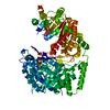
| ||||||||
|---|---|---|---|---|---|---|---|---|---|
| 1 |
| ||||||||
| Unit cell |
|
-Components
| #1: Protein | Mass: 88188.961 Da / Num. of mol.: 1 / Mutation: K103A, K104A, E107A Source method: isolated from a genetically manipulated source Source: (gene. exp.) Candida albicans (yeast) / Strain: BWP17 / Gene: CaO19.10083, CaO19.2551, MET6 / Plasmid: pNIC28-Bsa4 / Production host:  Escherichia coli (E. coli) / Strain (production host): BL21(DE3) Escherichia coli (E. coli) / Strain (production host): BL21(DE3)References: UniProt: P82610, 5-methyltetrahydropteroyltriglutamate-homocysteine S-methyltransferase |
|---|---|
| #2: Chemical | ChemComp-ZN /   Mass: 65.409 Da / Num. of mol.: 1 / Source method: obtained synthetically / Formula: Zn Mass: 65.409 Da / Num. of mol.: 1 / Source method: obtained synthetically / Formula: Zn |
| #3: Water | ChemComp-HOH / Water Mass: 18.015 Da / Num. of mol.: 501 / Source method: isolated from a natural source / Formula: H2O Mass: 18.015 Da / Num. of mol.: 501 / Source method: isolated from a natural source / Formula: H2O |
-Experimental details
-Experiment
| Experiment | Method: X-RAY DIFFRACTION / Number of used crystals: 1 |
|---|
- Sample preparation
| Crystal | Density Matthews: 2.25 Å3/Da / Density % sol: 45.44 % |
|---|---|
| Crystal grow | Temperature: 293 K / Method: vapor diffusion, hanging drop / pH: 7.4 Details: 50 mM NaI, 27% (w/v) PEG 3350, pH 7.4, VAPOR DIFFUSION, HANGING DROP, temperature 293K |
-Data collection
| Diffraction | Mean temperature: 100 K | |||||||||||||||||||||||||||||||||||||||||||||||||||||||||||||||||||||||||||||||||||||||||||||||||||||||||
|---|---|---|---|---|---|---|---|---|---|---|---|---|---|---|---|---|---|---|---|---|---|---|---|---|---|---|---|---|---|---|---|---|---|---|---|---|---|---|---|---|---|---|---|---|---|---|---|---|---|---|---|---|---|---|---|---|---|---|---|---|---|---|---|---|---|---|---|---|---|---|---|---|---|---|---|---|---|---|---|---|---|---|---|---|---|---|---|---|---|---|---|---|---|---|---|---|---|---|---|---|---|---|---|---|---|---|
| Diffraction source | Source: SYNCHROTRON / Site: ALS  / Beamline: 5.0.3 / Wavelength: 0.9765 Å / Beamline: 5.0.3 / Wavelength: 0.9765 Å | |||||||||||||||||||||||||||||||||||||||||||||||||||||||||||||||||||||||||||||||||||||||||||||||||||||||||
| Detector | Type: ADSC QUANTUM 315r / Detector: CCD / Date: Jul 12, 2010 / Details: Asymmetric cut single crystal Si (220) | |||||||||||||||||||||||||||||||||||||||||||||||||||||||||||||||||||||||||||||||||||||||||||||||||||||||||
| Radiation | Monochromator: Asymmetric cut single crystal Si (220) / Protocol: SINGLE WAVELENGTH / Monochromatic (M) / Laue (L): M / Scattering type: x-ray | |||||||||||||||||||||||||||||||||||||||||||||||||||||||||||||||||||||||||||||||||||||||||||||||||||||||||
| Radiation wavelength | Wavelength: 0.9765 Å / Relative weight: 1 | |||||||||||||||||||||||||||||||||||||||||||||||||||||||||||||||||||||||||||||||||||||||||||||||||||||||||
| Reflection | Resolution: 1.98→50 Å / Num. all: 54885 / Num. obs: 54885 / % possible obs: 100 % / Redundancy: 10 % / Rmerge(I) obs: 0.084 / Net I/σ(I): 10.1 | |||||||||||||||||||||||||||||||||||||||||||||||||||||||||||||||||||||||||||||||||||||||||||||||||||||||||
| Reflection shell |
|
- Processing
| Software |
| ||||||||||||||||||||||||||||||||||||||||||||||||||||||||||||||||||||||||||||||||||||||||||||||||||||||||||||||||||||||||||||||||||||||||||||||||||||||||||||||||||||||||||||||||||||||||||||||||||||||||
|---|---|---|---|---|---|---|---|---|---|---|---|---|---|---|---|---|---|---|---|---|---|---|---|---|---|---|---|---|---|---|---|---|---|---|---|---|---|---|---|---|---|---|---|---|---|---|---|---|---|---|---|---|---|---|---|---|---|---|---|---|---|---|---|---|---|---|---|---|---|---|---|---|---|---|---|---|---|---|---|---|---|---|---|---|---|---|---|---|---|---|---|---|---|---|---|---|---|---|---|---|---|---|---|---|---|---|---|---|---|---|---|---|---|---|---|---|---|---|---|---|---|---|---|---|---|---|---|---|---|---|---|---|---|---|---|---|---|---|---|---|---|---|---|---|---|---|---|---|---|---|---|---|---|---|---|---|---|---|---|---|---|---|---|---|---|---|---|---|---|---|---|---|---|---|---|---|---|---|---|---|---|---|---|---|---|---|---|---|---|---|---|---|---|---|---|---|---|---|---|---|---|
| Refinement | Method to determine structure: MOLECULAR REPLACEMENT Starting model: PDB entry 3PPC Resolution: 1.98→49.609 Å / Occupancy max: 1 / Occupancy min: 1 / SU ML: 0.22 / σ(F): 0 / Phase error: 21.49 / Stereochemistry target values: ML
| ||||||||||||||||||||||||||||||||||||||||||||||||||||||||||||||||||||||||||||||||||||||||||||||||||||||||||||||||||||||||||||||||||||||||||||||||||||||||||||||||||||||||||||||||||||||||||||||||||||||||
| Solvent computation | Shrinkage radii: 1.06 Å / VDW probe radii: 1.3 Å / Solvent model: FLAT BULK SOLVENT MODEL / Bsol: 43.335 Å2 / ksol: 0.283 e/Å3 | ||||||||||||||||||||||||||||||||||||||||||||||||||||||||||||||||||||||||||||||||||||||||||||||||||||||||||||||||||||||||||||||||||||||||||||||||||||||||||||||||||||||||||||||||||||||||||||||||||||||||
| Displacement parameters |
| ||||||||||||||||||||||||||||||||||||||||||||||||||||||||||||||||||||||||||||||||||||||||||||||||||||||||||||||||||||||||||||||||||||||||||||||||||||||||||||||||||||||||||||||||||||||||||||||||||||||||
| Refinement step | Cycle: LAST / Resolution: 1.98→49.609 Å
| ||||||||||||||||||||||||||||||||||||||||||||||||||||||||||||||||||||||||||||||||||||||||||||||||||||||||||||||||||||||||||||||||||||||||||||||||||||||||||||||||||||||||||||||||||||||||||||||||||||||||
| Refine LS restraints |
| ||||||||||||||||||||||||||||||||||||||||||||||||||||||||||||||||||||||||||||||||||||||||||||||||||||||||||||||||||||||||||||||||||||||||||||||||||||||||||||||||||||||||||||||||||||||||||||||||||||||||
| LS refinement shell |
| ||||||||||||||||||||||||||||||||||||||||||||||||||||||||||||||||||||||||||||||||||||||||||||||||||||||||||||||||||||||||||||||||||||||||||||||||||||||||||||||||||||||||||||||||||||||||||||||||||||||||
| Refinement TLS params. | Method: refined / Refine-ID: X-RAY DIFFRACTION
| ||||||||||||||||||||||||||||||||||||||||||||||||||||||||||||||||||||||||||||||||||||||||||||||||||||||||||||||||||||||||||||||||||||||||||||||||||||||||||||||||||||||||||||||||||||||||||||||||||||||||
| Refinement TLS group |
|
