 Movie
Movie Controller


+ Open data

- Basic information
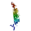
| Entry | Database: PDB / ID: 3ppe | ||||||
|---|---|---|---|---|---|---|---|
| Title | Crystal structure of chicken VE-cadherin EC1-2 | ||||||
 Components Components | Vascular endothelial cadherin | ||||||
 Keywords Keywords |  CELL ADHESION / extracellular cadherin (EC) domain / beta barrel / Ig-domain like / domain swapped dimer interface / calcium dependent cell-cell adhesion / cell surface CELL ADHESION / extracellular cadherin (EC) domain / beta barrel / Ig-domain like / domain swapped dimer interface / calcium dependent cell-cell adhesion / cell surface | ||||||
| Function / homology |  Function and homology information Function and homology informationVEGFR2 mediated vascular permeability / Adherens junctions interactions / positive regulation of establishment of endothelial barrier / blood vessel maturation / blood vessel endothelial cell migration / cell-cell adhesion mediated by cadherin / cardiac epithelial to mesenchymal transition / protein localization to bicellular tight junction / regulation of vascular permeability / BMP receptor binding ...VEGFR2 mediated vascular permeability / Adherens junctions interactions / positive regulation of establishment of endothelial barrier / blood vessel maturation / blood vessel endothelial cell migration / cell-cell adhesion mediated by cadherin / cardiac epithelial to mesenchymal transition / protein localization to bicellular tight junction / regulation of vascular permeability / BMP receptor binding / cell-cell adhesion via plasma-membrane adhesion molecules / bicellular tight junction assembly / fibrinogen binding / calcium-dependent cell-cell adhesion via plasma membrane cell adhesion molecules / vascular endothelial growth factor receptor 2 binding / positive regulation of BMP signaling pathway / vasculature development / catenin complex / regulation of establishment of cell polarity / cell-cell junction assembly / adherens junction organization / negative regulation of microtubule polymerization / homophilic cell adhesion via plasma membrane adhesion molecules / bicellular tight junction / negative regulation of endothelial cell apoptotic process / positive regulation of protein dephosphorylation / protein tyrosine kinase binding / transforming growth factor beta receptor signaling pathway / positive regulation of protein-containing complex assembly / adherens junction / regulation of protein phosphorylation / cell morphogenesis / beta-catenin binding / intracellular calcium ion homeostasis / negative regulation of inflammatory response / positive regulation of angiogenesis / protein phosphatase binding / nuclear membrane / transmembrane transporter binding / membrane => GO:0016020 / positive regulation of cell migration / cadherin binding / negative regulation of cell population proliferation / external side of plasma membrane / calcium ion binding / positive regulation of gene expression / nucleoplasmSimilarity search - Function | ||||||
| Biological species |  Gallus gallus (chicken) Gallus gallus (chicken) | ||||||
| Method | X-RAY DIFFRACTION / SYNCHROTRON / MOLECULAR REPLACEMENT / Resolution: 2.1 Å | ||||||
 Authors Authors | Brasch, J. / Harrison, O.J. / Ahlsen, G. / Carnally, S.M. / Henderson, R.M. / Honig, B. / Shapiro, L.S. | ||||||
 Citation Citation | Journal: J.Mol.Biol. / Year: 2011 Title: Structure and binding mechanism of vascular endothelial cadherin: a divergent classical cadherin. Authors: Brasch, J. / Harrison, O.J. / Ahlsen, G. / Carnally, S.M. / Henderson, R.M. / Honig, B. / Shapiro, L. | ||||||
| History |
|
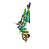
- Structure visualization
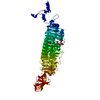
| Structure viewer | Molecule: MolmilJmol/JSmol |
|---|
- Downloads & links
-Download
| PDBx/mmCIF format | 3ppe.cif.gz | 109.3 KB | Display | PDBx/mmCIF format |
|---|---|---|---|---|
| PDB format | pdb3ppe.ent.gz | 83.6 KB | Display | PDB format |
| PDBx/mmJSON format | 3ppe.json.gz | Tree view | PDBx/mmJSON format | |
| Others |  Other downloads Other downloads |
-Validation report
| Arichive directory | https://data.pdbj.org/pub/pdb/validation_reports/pp/3ppeftp://data.pdbj.org/pub/pdb/validation_reports/pp/3ppe | HTTPS FTP |
|---|
-Related structure data
| Related structure data | 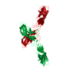 2a4eS S: Starting model for refinement |
|---|---|
| Similar structure data |


-Links
 PDBj
PDBj
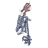
- Assembly
| Deposited unit | 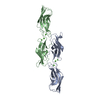
| ||||||||
|---|---|---|---|---|---|---|---|---|---|
| 1 |
| ||||||||
| Unit cell |
| ||||||||
| Components on special symmetry positions |
|
-Components
| #1: Protein | Mass: 22693.367 Da / Num. of mol.: 2 Fragment: chicken VE-cadherin domains EC1-EC2, UNP residues 41-243 Source method: isolated from a genetically manipulated source Source: (gene. exp.) Gallus gallus (chicken) / Plasmid: pSMT3 / Production host:  Escherichia coli (E. coli) / Strain (production host): Rosetta2DE3 pLysS / References: UniProt: Q8AYD0 Escherichia coli (E. coli) / Strain (production host): Rosetta2DE3 pLysS / References: UniProt: Q8AYD0#2: Chemical | ChemComp-CA /   Mass: 40.078 Da / Num. of mol.: 6 / Source method: obtained synthetically / Formula: Ca Mass: 40.078 Da / Num. of mol.: 6 / Source method: obtained synthetically / Formula: Ca#3: Water | ChemComp-HOH / | Water Mass: 18.015 Da / Num. of mol.: 587 / Source method: isolated from a natural source / Formula: H2O Mass: 18.015 Da / Num. of mol.: 587 / Source method: isolated from a natural source / Formula: H2O |
|---|
-Experimental details
-Experiment
| Experiment | Method: X-RAY DIFFRACTION / Number of used crystals: 1 |
|---|
- Sample preparation
| Crystal | Density Matthews: 2.92 Å3/Da / Density % sol: 57.84 % |
|---|---|
| Crystal grow | Temperature: 293.15 K / Method: vapor diffusion, hanging drop / pH: 6.5 Details: 18% (w/v) PEG 8,000, 200mM Calcium acetate, 100mM sodium cacodylate pH6.5., VAPOR DIFFUSION, HANGING DROP, temperature 293.15K |
-Data collection
| Diffraction | Mean temperature: 100 K |
|---|---|
| Diffraction source | Source: SYNCHROTRON / Site: NSLS  / Beamline: X4C / Wavelength: 0.97885 Å / Beamline: X4C / Wavelength: 0.97885 Å |
| Detector | Type: MAR scanner 345 mm plate / Detector: IMAGE PLATE / Date: Apr 9, 2009 |
| Radiation | Monochromator: Bent single Si(111) crystal (horizontal focusing and deflection) Protocol: SINGLE WAVELENGTH / Monochromatic (M) / Laue (L): M / Scattering type: x-ray |
| Radiation wavelength | Wavelength: 0.97885 Å / Relative weight: 1 |
| Reflection | Resolution: 2.1→80 Å / Num. obs: 31991 / % possible obs: 100 % / Redundancy: 14.1 % |
| Reflection shell | Resolution: 2.1→2.18 Å / % possible all: 100 |
- Processing
| Software |
| ||||||||||||||||||||||||||||||||||||||||||||||||||||||||||||||||||||||||||||||||||||||||||||||||||||||||||||||||||||||||||||||||||||||||||||||||||||||||||||||||||||||||||
|---|---|---|---|---|---|---|---|---|---|---|---|---|---|---|---|---|---|---|---|---|---|---|---|---|---|---|---|---|---|---|---|---|---|---|---|---|---|---|---|---|---|---|---|---|---|---|---|---|---|---|---|---|---|---|---|---|---|---|---|---|---|---|---|---|---|---|---|---|---|---|---|---|---|---|---|---|---|---|---|---|---|---|---|---|---|---|---|---|---|---|---|---|---|---|---|---|---|---|---|---|---|---|---|---|---|---|---|---|---|---|---|---|---|---|---|---|---|---|---|---|---|---|---|---|---|---|---|---|---|---|---|---|---|---|---|---|---|---|---|---|---|---|---|---|---|---|---|---|---|---|---|---|---|---|---|---|---|---|---|---|---|---|---|---|---|---|---|---|---|---|---|
| Refinement | Method to determine structure: MOLECULAR REPLACEMENT Starting model: 2A4E Resolution: 2.1→72.74 Å / Cor.coef. Fo:Fc: 0.945 / Cor.coef. Fo:Fc free: 0.904 / Cross valid method: THROUGHOUT / ESU R: 0.199 / ESU R Free: 0.184 / Stereochemistry target values: MAXIMUM LIKELIHOOD / Details: HYDROGENS HAVE BEEN ADDED IN THE RIDING POSITIONS
| ||||||||||||||||||||||||||||||||||||||||||||||||||||||||||||||||||||||||||||||||||||||||||||||||||||||||||||||||||||||||||||||||||||||||||||||||||||||||||||||||||||||||||
| Solvent computation | Ion probe radii: 0.8 Å / Shrinkage radii: 0.8 Å / VDW probe radii: 1.4 Å / Solvent model: MASK | ||||||||||||||||||||||||||||||||||||||||||||||||||||||||||||||||||||||||||||||||||||||||||||||||||||||||||||||||||||||||||||||||||||||||||||||||||||||||||||||||||||||||||
| Displacement parameters | Biso mean: 21.211 Å2
| ||||||||||||||||||||||||||||||||||||||||||||||||||||||||||||||||||||||||||||||||||||||||||||||||||||||||||||||||||||||||||||||||||||||||||||||||||||||||||||||||||||||||||
| Refinement step | Cycle: LAST / Resolution: 2.1→72.74 Å
| ||||||||||||||||||||||||||||||||||||||||||||||||||||||||||||||||||||||||||||||||||||||||||||||||||||||||||||||||||||||||||||||||||||||||||||||||||||||||||||||||||||||||||
| Refine LS restraints |
| ||||||||||||||||||||||||||||||||||||||||||||||||||||||||||||||||||||||||||||||||||||||||||||||||||||||||||||||||||||||||||||||||||||||||||||||||||||||||||||||||||||||||||
| LS refinement shell | Resolution: 2.101→2.155 Å / Total num. of bins used: 20
|
