 Movie
Movie Controller


+ Open data

- Basic information
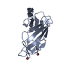
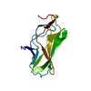
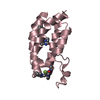
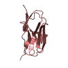
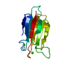
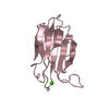
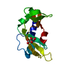
| Entry | Database: PDB / ID: 3pof | ||||||
|---|---|---|---|---|---|---|---|
| Title | Crystal structure of MASP-1 CUB2 domain bound to Ca2+ | ||||||
 Components Components | Mannan-binding lectin serine protease 1 | ||||||
 Keywords Keywords |  HYDROLASE / CUB domain / Ca2+ binding site / complement protein / lectin pathway of complement / MBL / MBP / ficolins / Bloodstream HYDROLASE / CUB domain / Ca2+ binding site / complement protein / lectin pathway of complement / MBL / MBP / ficolins / Bloodstream | ||||||
| Function / homology |  Function and homology information Function and homology informationScavenging by Class A Receptors / Ficolins bind to repetitive carbohydrate structures on the target cell surface / Lectin pathway of complement activation / Initial triggering of complement / complement activation, lectin pathway / complement activation / zymogen activation / Hydrolases; Acting on peptide bonds (peptidases); Serine endopeptidases / calcium-dependent protein binding / peptidase activity ...Scavenging by Class A Receptors / Ficolins bind to repetitive carbohydrate structures on the target cell surface / Lectin pathway of complement activation / Initial triggering of complement / complement activation, lectin pathway / complement activation / zymogen activation / Hydrolases; Acting on peptide bonds (peptidases); Serine endopeptidases / calcium-dependent protein binding / peptidase activity / serine-type endopeptidase activity / calcium ion binding / protein homodimerization activity / extracellular spaceSimilarity search - Function | ||||||
| Biological species |  Rattus norvegicus (Norway rat) Rattus norvegicus (Norway rat) | ||||||
| Method | X-RAY DIFFRACTION / SYNCHROTRON / MOLECULAR REPLACEMENT / Resolution: 1.501 Å | ||||||
 Authors Authors | Gingras, A.R. / Moody, P.C.E. / Wallis, R. | ||||||
 Citation Citation | Journal: Structure / Year: 2011 Title: Structural Basis of Mannan-Binding Lectin Recognition by Its Associated Serine Protease MASP-1: Implications for Complement Activation. Authors: Gingras, A.R. / Girija, U.V. / Keeble, A.H. / Panchal, R. / Mitchell, D.A. / Moody, P.C. / Wallis, R. | ||||||
| History |
|
- Structure visualization
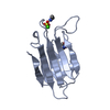
| Structure viewer | Molecule: MolmilJmol/JSmol |
|---|
- Downloads & links
-Download
| PDBx/mmCIF format | 3pof.cif.gz | 122.7 KB | Display | PDBx/mmCIF format |
|---|---|---|---|---|
| PDB format | pdb3pof.ent.gz | 95.4 KB | Display | PDB format |
| PDBx/mmJSON format | 3pof.json.gz | Tree view | PDBx/mmJSON format | |
| Others |  Other downloads Other downloads |
-Validation report
| Arichive directory | https://data.pdbj.org/pub/pdb/validation_reports/po/3pofftp://data.pdbj.org/pub/pdb/validation_reports/po/3pof | HTTPS FTP |
|---|
-Related structure data
| Related structure data | 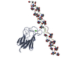 3pobC  3podC  3poeSC 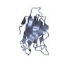 3pogC 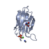 3poiC  3pojC  3ponC C: citing same article ( S: Starting model for refinement |
|---|---|
| Similar structure data |


-Links
 PDBj
PDBj


- Assembly
| Deposited unit | 
| |||||||||||||||
|---|---|---|---|---|---|---|---|---|---|---|---|---|---|---|---|---|
| 1 | 
| |||||||||||||||
| 2 | 
| |||||||||||||||
| Unit cell |
| |||||||||||||||
| Components on special symmetry positions |
|
-Components
| #1: Protein | Mass: 13091.511 Da / Num. of mol.: 2 / Fragment: MASP-1 CUB2 domain (UNP Residues 188-301) Source method: isolated from a genetically manipulated source Source: (gene. exp.) Rattus norvegicus (Norway rat) / Gene: Crarf, MASP-1, Masp1, Masp3 / Plasmid: pET28A / Production host:  Escherichia coli (E. coli) / Strain (production host): BL21(DE3) Escherichia coli (E. coli) / Strain (production host): BL21(DE3)References: UniProt: Q8CHN8, Hydrolases; Acting on peptide bonds (peptidases); Serine endopeptidases#2: Chemical |   Mass: 40.078 Da / Num. of mol.: 2 / Source method: obtained synthetically / Formula: Ca Mass: 40.078 Da / Num. of mol.: 2 / Source method: obtained synthetically / Formula: Ca#3: Chemical | Sulfate  Mass: 96.063 Da / Num. of mol.: 2 / Source method: obtained synthetically / Formula: SO4 Mass: 96.063 Da / Num. of mol.: 2 / Source method: obtained synthetically / Formula: SO4#4: Chemical | ChemComp-TRS / | Tris  Mass: 122.143 Da / Num. of mol.: 1 / Source method: obtained synthetically / Formula: C4H12NO3 / Comment: pH buffer*YM Mass: 122.143 Da / Num. of mol.: 1 / Source method: obtained synthetically / Formula: C4H12NO3 / Comment: pH buffer*YM#5: Water | ChemComp-HOH / | Water Mass: 18.015 Da / Num. of mol.: 251 / Source method: isolated from a natural source / Formula: H2O Mass: 18.015 Da / Num. of mol.: 251 / Source method: isolated from a natural source / Formula: H2O |
|---|
-Experimental details
-Experiment
| Experiment | Method: X-RAY DIFFRACTION / Number of used crystals: 1 |
|---|
- Sample preparation
| Crystal | Density Matthews: 3.23 Å3/Da / Density % sol: 61.88 % |
|---|---|
| Crystal grow | Temperature: 279 K / Method: vapor diffusion, sitting drop / pH: 8.5 Details: 1.5M lithium sulfate, 100mM tris buffer, pH 8.5, VAPOR DIFFUSION, SITTING DROP, temperature 279K |
-Data collection
| Diffraction | Mean temperature: 100 K |
|---|---|
| Diffraction source | Source: SYNCHROTRON / Site: Diamond  / Beamline: I03 / Wavelength: 0.9763 Å / Beamline: I03 / Wavelength: 0.9763 Å |
| Detector | Type: ADSC QUANTUM 315 / Detector: CCD / Date: Jul 3, 2010 |
| Radiation | Monochromator: multi-layer optics / Protocol: SINGLE WAVELENGTH / Monochromatic (M) / Laue (L): M / Scattering type: x-ray |
| Radiation wavelength | Wavelength: 0.9763 Å / Relative weight: 1 |
| Reflection | Resolution: 1.5→50 Å / Num. obs: 53908 / % possible obs: 99.6 % / Observed criterion σ(F): 0 / Observed criterion σ(I): 0 / Redundancy: 4.92 % / Rmerge(I) obs: 0.076 / Net I/σ(I): 18.5 |
| Reflection shell | Resolution: 1.5→1.59 Å / Redundancy: 4.92 % / Rmerge(I) obs: 0.331 / Mean I/σ(I) obs: 4.38 / Num. unique all: 42562 / % possible all: 99.7 |
- Processing
| Software |
| ||||||||||||||||||||||||||||||||||||||||||||||||||||||||||||||||||||||||||||||||||||||||||||||||||||||||||||||||||||||||||||||||||||||||||||||||||||||||||||||||||||||||||
|---|---|---|---|---|---|---|---|---|---|---|---|---|---|---|---|---|---|---|---|---|---|---|---|---|---|---|---|---|---|---|---|---|---|---|---|---|---|---|---|---|---|---|---|---|---|---|---|---|---|---|---|---|---|---|---|---|---|---|---|---|---|---|---|---|---|---|---|---|---|---|---|---|---|---|---|---|---|---|---|---|---|---|---|---|---|---|---|---|---|---|---|---|---|---|---|---|---|---|---|---|---|---|---|---|---|---|---|---|---|---|---|---|---|---|---|---|---|---|---|---|---|---|---|---|---|---|---|---|---|---|---|---|---|---|---|---|---|---|---|---|---|---|---|---|---|---|---|---|---|---|---|---|---|---|---|---|---|---|---|---|---|---|---|---|---|---|---|---|---|---|---|
| Refinement | Method to determine structure: MOLECULAR REPLACEMENT Starting model: PDB ENTRY 3POE Resolution: 1.501→41.01 Å / Cor.coef. Fo:Fc: 0.977 / Cor.coef. Fo:Fc free: 0.968 / SU B: 1.466 / SU ML: 0.025 / Cross valid method: THROUGHOUT / ESU R: 0.048 / ESU R Free: 0.047 / Stereochemistry target values: MAXIMUM LIKELIHOOD / Details: HYDROGENS HAVE BEEN ADDED IN THE RIDING POSITIONS
| ||||||||||||||||||||||||||||||||||||||||||||||||||||||||||||||||||||||||||||||||||||||||||||||||||||||||||||||||||||||||||||||||||||||||||||||||||||||||||||||||||||||||||
| Solvent computation | Ion probe radii: 0.8 Å / Shrinkage radii: 0.8 Å / VDW probe radii: 1.4 Å / Solvent model: MASK | ||||||||||||||||||||||||||||||||||||||||||||||||||||||||||||||||||||||||||||||||||||||||||||||||||||||||||||||||||||||||||||||||||||||||||||||||||||||||||||||||||||||||||
| Displacement parameters | Biso mean: 13.404 Å2 | ||||||||||||||||||||||||||||||||||||||||||||||||||||||||||||||||||||||||||||||||||||||||||||||||||||||||||||||||||||||||||||||||||||||||||||||||||||||||||||||||||||||||||
| Refinement step | Cycle: LAST / Resolution: 1.501→41.01 Å
| ||||||||||||||||||||||||||||||||||||||||||||||||||||||||||||||||||||||||||||||||||||||||||||||||||||||||||||||||||||||||||||||||||||||||||||||||||||||||||||||||||||||||||
| Refine LS restraints |
| ||||||||||||||||||||||||||||||||||||||||||||||||||||||||||||||||||||||||||||||||||||||||||||||||||||||||||||||||||||||||||||||||||||||||||||||||||||||||||||||||||||||||||
| LS refinement shell | Resolution: 1.501→1.54 Å / Total num. of bins used: 20
|
