 Movie
Movie Controller

[English] 日本語
 Yorodumi
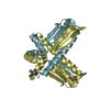
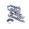
- PDB-3pjt: Structure of Pseudomonas fluorescence LapD EAL domain complexed w... -
+ Open data

- Basic information
| Entry | Database: PDB / ID: 3pjt | ||||||
|---|---|---|---|---|---|---|---|
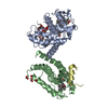
| Title | Structure of Pseudomonas fluorescence LapD EAL domain complexed with c-di-GMP, C2221 | ||||||
 Components Components | Cyclic dimeric GMP binding protein | ||||||
 Keywords Keywords |  LYASE / Tim Barrel / c-di-GMP receptor LYASE / Tim Barrel / c-di-GMP receptor | ||||||
| Function / homology |  Function and homology informationnucleotide binding / signal transduction / membrane / identical protein binding Function and homology informationnucleotide binding / signal transduction / membrane / identical protein bindingSimilarity search - Function | ||||||
| Biological species |  Pseudomonas fluorescens (bacteria) Pseudomonas fluorescens (bacteria) | ||||||
| Method | X-RAY DIFFRACTION / SYNCHROTRON / MOLECULAR REPLACEMENT / Resolution: 2.5154 Å | ||||||
 Authors Authors | Sondermann, H. / Navarro, M.V.A.S. / Krasteva, P. | ||||||
 Citation Citation | Journal: Plos Biol. / Year: 2011 Title: Structural Basis for c-di-GMP-Mediated Inside-Out Signaling Controlling Periplasmic Proteolysis. Authors: Navarro, M.V. / Newell, P.D. / Krasteva, P.V. / Chatterjee, D. / Madden, D.R. / O'Toole, G.A. / Sondermann, H. | ||||||
| History |
|
- Structure visualization
| Structure viewer | Molecule: MolmilJmol/JSmol |
|---|
- Downloads & links
-Download
| PDBx/mmCIF format | 3pjt.cif.gz | 112.7 KB | Display | PDBx/mmCIF format |
|---|---|---|---|---|
| PDB format | pdb3pjt.ent.gz | 87.5 KB | Display | PDB format |
| PDBx/mmJSON format | 3pjt.json.gz | Tree view | PDBx/mmJSON format | |
| Others |  Other downloads Other downloads |
-Validation report
| Arichive directory | https://data.pdbj.org/pub/pdb/validation_reports/pj/3pjtftp://data.pdbj.org/pub/pdb/validation_reports/pj/3pjt | HTTPS FTP |
|---|
-Related structure data


-Links
 PDBj
PDBj


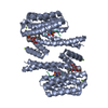
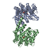
- Assembly
| Deposited unit | 
| ||||||||
|---|---|---|---|---|---|---|---|---|---|
| 1 |
| ||||||||
| Unit cell |
|
-Components
| #1: Protein | Mass: 28293.305 Da / Num. of mol.: 2 / Fragment: UNP Residues 400-648 Source method: isolated from a genetically manipulated source Source: (gene. exp.) Pseudomonas fluorescens (bacteria) / Strain: Pf0-1 / Gene: lapD, Pfl01_0131 / Production host: Escherichia coli (E. coli) / References: UniProt: Q3KK31#2: Chemical | Cyclic di-GMP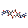  Mass: 690.411 Da / Num. of mol.: 2 / Source method: obtained synthetically / Formula: C20H24N10O14P2 Mass: 690.411 Da / Num. of mol.: 2 / Source method: obtained synthetically / Formula: C20H24N10O14P2#3: Water | ChemComp-HOH / | Water Mass: 18.015 Da / Num. of mol.: 115 / Source method: isolated from a natural source / Formula: H2O Mass: 18.015 Da / Num. of mol.: 115 / Source method: isolated from a natural source / Formula: H2O |
|---|
-Experimental details
-Experiment
| Experiment | Method: X-RAY DIFFRACTION / Number of used crystals: 1 |
|---|
- Sample preparation
| Crystal | Density Matthews: 2.67 Å3/Da / Density % sol: 53.94 % |
|---|---|
| Crystal grow | Temperature: 293 K / Method: vapor diffusion, hanging drop / pH: 5.5 Details: 0.1 M Bis-Tris, 0.2 M Ammonium sulfate, 24% PEG 3350, pH 5.5, VAPOR DIFFUSION, HANGING DROP, temperature 293K |
-Data collection
| Diffraction | Mean temperature: 100 K |
|---|---|
| Diffraction source | Source: SYNCHROTRON / Site: CHESS  / Beamline: A1 / Wavelength: 0.9769 Å / Beamline: A1 / Wavelength: 0.9769 Å |
| Detector | Type: ADSC QUANTUM 210 / Detector: CCD / Date: Feb 1, 2010 |
| Radiation | Monochromator: Si 111 CHANNEL / Protocol: SINGLE WAVELENGTH / Monochromatic (M) / Laue (L): M / Scattering type: x-ray |
| Radiation wavelength | Wavelength: 0.9769 Å / Relative weight: 1 |
| Reflection | Resolution: 2.5→50 Å / Num. all: 21133 / Num. obs: 20943 / % possible obs: 99.1 % / Observed criterion σ(F): 1 / Observed criterion σ(I): 1 |
| Reflection shell | Resolution: 2.5→2.59 Å / % possible all: 94.4 |
- Processing
| Software |
| |||||||||||||||||||||||||||||||||||||||||||||||||||||||||||||||||||||||||||||||||||||||||||||||||||||||||
|---|---|---|---|---|---|---|---|---|---|---|---|---|---|---|---|---|---|---|---|---|---|---|---|---|---|---|---|---|---|---|---|---|---|---|---|---|---|---|---|---|---|---|---|---|---|---|---|---|---|---|---|---|---|---|---|---|---|---|---|---|---|---|---|---|---|---|---|---|---|---|---|---|---|---|---|---|---|---|---|---|---|---|---|---|---|---|---|---|---|---|---|---|---|---|---|---|---|---|---|---|---|---|---|---|---|---|
| Refinement | Method to determine structure: MOLECULAR REPLACEMENT / Resolution: 2.5154→34.138 Å / SU ML: 0.37 / σ(F): 0.2 / Phase error: 23.8 / Stereochemistry target values: ML
| |||||||||||||||||||||||||||||||||||||||||||||||||||||||||||||||||||||||||||||||||||||||||||||||||||||||||
| Solvent computation | Shrinkage radii: 0.9 Å / VDW probe radii: 1.11 Å / Solvent model: FLAT BULK SOLVENT MODEL / Bsol: 41.309 Å2 / ksol: 0.379 e/Å3 | |||||||||||||||||||||||||||||||||||||||||||||||||||||||||||||||||||||||||||||||||||||||||||||||||||||||||
| Displacement parameters |
| |||||||||||||||||||||||||||||||||||||||||||||||||||||||||||||||||||||||||||||||||||||||||||||||||||||||||
| Refinement step | Cycle: LAST / Resolution: 2.5154→34.138 Å
| |||||||||||||||||||||||||||||||||||||||||||||||||||||||||||||||||||||||||||||||||||||||||||||||||||||||||
| Refine LS restraints |
| |||||||||||||||||||||||||||||||||||||||||||||||||||||||||||||||||||||||||||||||||||||||||||||||||||||||||
| LS refinement shell |
|
