 Movie
Movie Controller

[English] 日本語
 Yorodumi
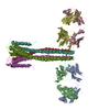
- PDB-3pjs: Mechanism of Activation Gating in the Full-Length KcsA K+ Channel -
+ Open data

- Basic information
| Entry | Database: PDB / ID: 3pjs | ||||||
|---|---|---|---|---|---|---|---|
| Title | Mechanism of Activation Gating in the Full-Length KcsA K+ Channel | ||||||
 Components Components |
| ||||||
 Keywords Keywords |  TRANSPORT PROTEIN / Ion channel / conducts K+ ions / Cell membrane TRANSPORT PROTEIN / Ion channel / conducts K+ ions / Cell membrane | ||||||
| Function / homology |  Function and homology information Function and homology informationmonoatomic ion transmembrane transport / identical protein binding / plasma membraneSimilarity search - Function | ||||||
| Biological species |  Mus musculus (house mouse) Mus musculus (house mouse) Streptomyces lividans (bacteria) Streptomyces lividans (bacteria) | ||||||
| Method | X-RAY DIFFRACTION / SYNCHROTRON / MOLECULAR REPLACEMENT / Resolution: 3.8 Å | ||||||
 Authors Authors | Uysal, S. / Cuello, L.G. / Kossiakoff, A. / Perozo, E. | ||||||
 Citation Citation | Journal: Proc.Natl.Acad.Sci.USA / Year: 2011 Title: Mechanism of activation gating in the full-length KcsA K+ channel. Authors: Uysal, S. / Cuello, L.G. / Cortes, D.M. / Koide, S. / Kossiakoff, A.A. / Perozo, E. | ||||||
| History |
|
- Structure visualization
| Structure viewer | Molecule: MolmilJmol/JSmol |
|---|
- Downloads & links
-Download
| PDBx/mmCIF format | 3pjs.cif.gz | 249.2 KB | Display | PDBx/mmCIF format |
|---|---|---|---|---|
| PDB format | pdb3pjs.ent.gz | 208.7 KB | Display | PDB format |
| PDBx/mmJSON format | 3pjs.json.gz | Tree view | PDBx/mmJSON format | |
| Others |  Other downloads Other downloads |
-Validation report
| Arichive directory | https://data.pdbj.org/pub/pdb/validation_reports/pj/3pjsftp://data.pdbj.org/pub/pdb/validation_reports/pj/3pjs | HTTPS FTP |
|---|
-Related structure data
| Similar structure data |
|---|


-Links
 PDBj
PDBj


- Assembly
| Deposited unit | 
| ||||||||
|---|---|---|---|---|---|---|---|---|---|
| 1 |
| ||||||||
| Unit cell |
|
-Components
| #1: Antibody | Fragment antigen-binding Mass: 23562.074 Da / Num. of mol.: 2 Source method: isolated from a genetically manipulated source Source: (gene. exp.) Mus musculus (house mouse) / Production host: Escherichia coli (E. coli)#2: Antibody | Fragment antigen-bindingMass: 23905.643 Da / Num. of mol.: 2 Source method: isolated from a genetically manipulated source Source: (gene. exp.) Mus musculus (house mouse) / Production host: Escherichia coli (E. coli)#3: Protein | / KcsAMass: 18509.361 Da / Num. of mol.: 4 Source method: isolated from a genetically manipulated source Source: (gene. exp.) Streptomyces lividans (bacteria) / Gene: kcsA, skc1 / Production host: Escherichia coli (E. coli) / References: UniProt: P0A334 |
|---|
-Experimental details
-Experiment
| Experiment | Method: X-RAY DIFFRACTION / Number of used crystals: 1 |
|---|
- Sample preparation
| Crystal | Density Matthews: 5.71 Å3/Da / Density % sol: 78.47 % |
|---|---|
| Crystal grow | Temperature: 293 K / Method: vapor diffusion, hanging drop / pH: 7.5 Details: 0.2M Na/K phosphate, 0.1M Bis-Tris propane pH 7.5, 10% PEG3350, VAPOR DIFFUSION, HANGING DROP, temperature 293K |
-Data collection
| Diffraction | Mean temperature: 100 K |
|---|---|
| Diffraction source | Source: SYNCHROTRON / Site: APS  / Beamline: 23-ID-D / Wavelength: 0.9 / Beamline: 23-ID-D / Wavelength: 0.9 |
| Detector | Type: ADSC QUANTUM 315 / Detector: CCD / Date: Nov 30, 2007 |
| Radiation | Monochromator: double crystal / Protocol: SINGLE WAVELENGTH / Monochromatic (M) / Laue (L): M / Scattering type: x-ray |
| Radiation wavelength | Wavelength: 0.9 Å / Relative weight: 1 |
| Reflection | Resolution: 3.8→50 Å / Num. all: 35572 / Num. obs: 29324 / % possible obs: 82.4 % / Observed criterion σ(F): 1.6 / Observed criterion σ(I): 1.6 |
| Reflection shell | Highest resolution: 3.8 Å / % possible all: 93.9 |
- Processing
| Software |
| |||||||||||||||||||||||||
|---|---|---|---|---|---|---|---|---|---|---|---|---|---|---|---|---|---|---|---|---|---|---|---|---|---|---|
| Refinement | Method to determine structure: MOLECULAR REPLACEMENT / Resolution: 3.8→40 Å / Occupancy max: 1 / Occupancy min: 1 / σ(F): 0 / Stereochemistry target values: Engh & Huber
| |||||||||||||||||||||||||
| Solvent computation | Bsol: 124.786 Å2 | |||||||||||||||||||||||||
| Displacement parameters | Biso max: 222.19 Å2 / Biso mean: 177.8122 Å2 / Biso min: 30.56 Å2
| |||||||||||||||||||||||||
| Refinement step | Cycle: LAST / Resolution: 3.8→40 Å
| |||||||||||||||||||||||||
| Refine LS restraints |
| |||||||||||||||||||||||||
| Xplor file | Serial no: 1 / Param file: CNS_TOPPAR:protein_rep.param |
