 Movie
Movie Controller


+ Open data

- Basic information
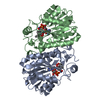
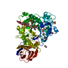
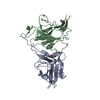
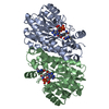
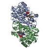
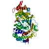
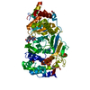
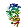
| Entry | Database: PDB / ID: 3pjf | ||||||
|---|---|---|---|---|---|---|---|
| Title | Structure of ENR G93V mutant-NAD+-triclosan complex | ||||||
 Components Components | Enoyl-[acyl-carrier-protein] reductase [NADH] | ||||||
 Keywords Keywords | OXIDOREDUCTASE/OXIDOREDUCTASE INHIBITOR /  ANTIBIOTIC RESISTANCE / FATTY ACID BIOSYNTHESIS / INNER MEMBRANE / LIPID SYNTHESIS / MEMBRANE / NAD / OXIDOREDUCTASE-OXIDOREDUCTASE INHIBITOR complex ANTIBIOTIC RESISTANCE / FATTY ACID BIOSYNTHESIS / INNER MEMBRANE / LIPID SYNTHESIS / MEMBRANE / NAD / OXIDOREDUCTASE-OXIDOREDUCTASE INHIBITOR complex | ||||||
| Function / homology |  Function and homology information Function and homology informationenoyl-[acyl-carrier-protein] reductase activity (NAD(P)H) / NADH binding / biotin biosynthetic process / fatty acid elongation / enoyl-[acyl-carrier-protein] reductase (NADH) / enoyl-[acyl-carrier-protein] reductase (NADH) activity / lipid biosynthetic process / catalytic complex / protein homotetramerization / response to antibiotic ...enoyl-[acyl-carrier-protein] reductase activity (NAD(P)H) / NADH binding / biotin biosynthetic process / fatty acid elongation / enoyl-[acyl-carrier-protein] reductase (NADH) / enoyl-[acyl-carrier-protein] reductase (NADH) activity / lipid biosynthetic process / catalytic complex / protein homotetramerization / response to antibiotic / protein-containing complex / membrane / identical protein binding / cytosolSimilarity search - Function | ||||||
| Biological species |  Escherichia coli (E. coli) Escherichia coli (E. coli) | ||||||
| Method | X-RAY DIFFRACTION / SYNCHROTRON / MOLECULAR REPLACEMENT / Resolution: 1.9 Å | ||||||
 Authors Authors | Kim, H.T. / Shin, D.G. / Chang, H.J. | ||||||
 Citation Citation | Journal: J.Struct.Biol. / Year: 2011 Title: Structural basis of triclosan resistance Authors: Jiten Singh, N. / Shin, D.G. / Lee, H.M. / Kim, H.T. / Chang, H.J. / Cho, J.M. / Kim, K.S. / Ro, S. | ||||||
| History |
|
- Structure visualization
| Structure viewer | Molecule: MolmilJmol/JSmol |
|---|
- Downloads & links
-Download
| PDBx/mmCIF format | 3pjf.cif.gz | 114.3 KB | Display | PDBx/mmCIF format |
|---|---|---|---|---|
| PDB format | pdb3pjf.ent.gz | 87.7 KB | Display | PDB format |
| PDBx/mmJSON format | 3pjf.json.gz | Tree view | PDBx/mmJSON format | |
| Others |  Other downloads Other downloads |
-Validation report
| Arichive directory | https://data.pdbj.org/pub/pdb/validation_reports/pj/3pjfftp://data.pdbj.org/pub/pdb/validation_reports/pj/3pjf | HTTPS FTP |
|---|
-Related structure data
| Related structure data |  3pjdC 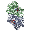 3pjeC 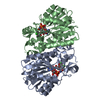 1c14S C: citing same article ( S: Starting model for refinement |
|---|---|
| Similar structure data |


-Links
 PDBj
PDBj

- Assembly
| Deposited unit | 
| ||||||||
|---|---|---|---|---|---|---|---|---|---|
| 1 |
| ||||||||
| Unit cell |
|
-Components
| #1: Protein | Mass: 29006.146 Da / Num. of mol.: 2 / Mutation: G93V Source method: isolated from a genetically manipulated source Source: (gene. exp.) Escherichia coli (E. coli) / Strain: K12 / Gene: ENVM, fabI / Plasmid: modified pET21b / Production host: Escherichia coli (E. coli) / Strain (production host): BL21(DE3)References: UniProt: P0AEK4, enoyl-[acyl-carrier-protein] reductase (NADH) #2: Chemical | Nicotinamide adenine dinucleotide  Mass: 663.425 Da / Num. of mol.: 2 / Source method: obtained synthetically / Formula: C21H27N7O14P2 / Comment: NAD*YM Mass: 663.425 Da / Num. of mol.: 2 / Source method: obtained synthetically / Formula: C21H27N7O14P2 / Comment: NAD*YM#3: Chemical | Triclosan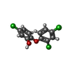  Mass: 289.542 Da / Num. of mol.: 2 / Source method: obtained synthetically / Formula: C12H7Cl3O2 / Comment: antifungal, antibiotic, detergent*YM Mass: 289.542 Da / Num. of mol.: 2 / Source method: obtained synthetically / Formula: C12H7Cl3O2 / Comment: antifungal, antibiotic, detergent*YM#4: Water | ChemComp-HOH / | Water Mass: 18.015 Da / Num. of mol.: 216 / Source method: isolated from a natural source / Formula: H2O Mass: 18.015 Da / Num. of mol.: 216 / Source method: isolated from a natural source / Formula: H2O |
|---|
-Experimental details
-Experiment
| Experiment | Method: X-RAY DIFFRACTION / Number of used crystals: 1 |
|---|
- Sample preparation
| Crystal | Density Matthews: 2.54 Å3/Da / Density % sol: 51.59 % |
|---|---|
| Crystal grow | Temperature: 295 K / Method: vapor diffusion, hanging drop / pH: 8.5 Details: 0.5M AMMONIUM CITRATE/AMMONIUM HYDROXIDE, 15% PEG8K, pH 8.50, VAPOR DIFFUSION, HANGING DROP, temperature 295K |
-Data collection
| Diffraction | Mean temperature: 100 K | |||||||||
|---|---|---|---|---|---|---|---|---|---|---|
| Diffraction source | Source: SYNCHROTRON / Site: PAL/PLS  / Beamline: 4A / Wavelength: 1 / Wavelength: 1 Å / Beamline: 4A / Wavelength: 1 / Wavelength: 1 Å | |||||||||
| Detector | Type: ADSC QUANTUM 210r / Detector: CCD / Date: Mar 11, 2007 | |||||||||
| Radiation | Monochromator: SAGITALLY FOCUSED SI(111) / Protocol: SINGLE WAVELENGTH / Monochromatic (M) / Laue (L): M / Scattering type: x-ray | |||||||||
| Radiation wavelength |
| |||||||||
| Reflection | Resolution: 1.9→50 Å / Num. obs: 46610 / % possible obs: 94.7 % / Observed criterion σ(F): 1 / Observed criterion σ(I): 1 / Redundancy: 11.4 % / Biso Wilson estimate: 17 Å2 / Rmerge(I) obs: 0.083 / Rsym value: 0.076 | |||||||||
| Reflection shell | Resolution: 1.9→1.97 Å / Redundancy: 6.9 % / Rmerge(I) obs: 0.276 / Mean I/σ(I) obs: 4.43 / Num. unique all: 4310 / Rsym value: 0.306 / % possible all: 90.1 |
- Processing
| Software |
| ||||||||||||||||||||||||||||||||||||||||||||||||||||||||||||||||||||||||||||||||
|---|---|---|---|---|---|---|---|---|---|---|---|---|---|---|---|---|---|---|---|---|---|---|---|---|---|---|---|---|---|---|---|---|---|---|---|---|---|---|---|---|---|---|---|---|---|---|---|---|---|---|---|---|---|---|---|---|---|---|---|---|---|---|---|---|---|---|---|---|---|---|---|---|---|---|---|---|---|---|---|---|---|
| Refinement | Method to determine structure: MOLECULAR REPLACEMENT Starting model: 1C14 Resolution: 1.9→50 Å / Rfactor Rfree error: 0.005 / Data cutoff high absF: 50577.18 / Data cutoff low absF: 0 / Isotropic thermal model: RESTRAINED / Cross valid method: THROUGHOUT / σ(F): 0 / σ(I): 0 / Stereochemistry target values: Engh & Huber
| ||||||||||||||||||||||||||||||||||||||||||||||||||||||||||||||||||||||||||||||||
| Solvent computation | Solvent model: FLAT MODEL / Bsol: 43.26 Å2 / ksol: 0.37 e/Å3 | ||||||||||||||||||||||||||||||||||||||||||||||||||||||||||||||||||||||||||||||||
| Displacement parameters | Biso mean: 28.3 Å2
| ||||||||||||||||||||||||||||||||||||||||||||||||||||||||||||||||||||||||||||||||
| Refine analyze |
| ||||||||||||||||||||||||||||||||||||||||||||||||||||||||||||||||||||||||||||||||
| Refinement step | Cycle: LAST / Resolution: 1.9→50 Å
| ||||||||||||||||||||||||||||||||||||||||||||||||||||||||||||||||||||||||||||||||
| Refine LS restraints |
| ||||||||||||||||||||||||||||||||||||||||||||||||||||||||||||||||||||||||||||||||
| LS refinement shell |
| ||||||||||||||||||||||||||||||||||||||||||||||||||||||||||||||||||||||||||||||||
| Xplor file |
|
