negative regulation of protein refolding / histone mRNA metabolic process / regulation of ATP-dependent activity / U2 snRNP binding / U7 snRNA binding / histone pre-mRNA DCP binding / U7 snRNP / histone pre-mRNA 3'end processing complex / SLBP independent Processing of Histone Pre-mRNAs / SLBP Dependent Processing of Replication-Dependent Histone Pre-mRNAs ...negative regulation of protein refolding / histone mRNA metabolic process / regulation of ATP-dependent activity / U2 snRNP binding / U7 snRNA binding / histone pre-mRNA DCP binding / U7 snRNP / histone pre-mRNA 3'end processing complex / SLBP independent Processing of Histone Pre-mRNAs / SLBP Dependent Processing of Replication-Dependent Histone Pre-mRNAs / import into nucleus / protein methylation / U12-type spliceosomal complex / methylosome / 7-methylguanosine cap hypermethylation / U1 snRNP binding / pICln-Sm protein complex / spliceosomal tri-snRNP complex / small nuclear ribonucleoprotein complex / P granule / SMN-Sm protein complex / U2-type spliceosomal complex / negative regulation of chaperone-mediated autophagy / telomerase RNA binding / U2-type precatalytic spliceosome / positive regulation of mRNA splicing, via spliceosome / telomerase holoenzyme complex / U2-type prespliceosome assembly / U2-type catalytic step 2 spliceosome / commitment complex / U4 snRNP / U2 snRNP / RNA Polymerase II Transcription Termination / U1 snRNP / U2-type prespliceosome / precatalytic spliceosome / termination of RNA polymerase II transcription / spliceosomal complex assembly / mRNA Splicing - Minor Pathway / regulation of RNA splicing / U5 snRNP / spliceosomal snRNP assembly / U1 snRNA binding / U4/U6 x U5 tri-snRNP complex / catalytic step 2 spliceosome / mRNA Splicing - Major Pathway / RNA splicing / spliceosomal complex / mRNA splicing, via spliceosome / snRNP Assembly / SARS-CoV-2 modulates host translation machinery / nuclear body / nuclear speck / ribonucleoprotein complex / mRNA binding / enzyme binding / DNA binding / RNA binding / extracellular exosome / nucleoplasm / identical protein binding / nucleus / cytosol / cytoplasm Similarity search - Function
snRNP70, RNA recognition motif / U1 small nuclear ribonucleoprotein of 70kDa N-terminal / U1 small nuclear ribonucleoprotein of 70kDa MW N terminal / U1 small nuclear ribonucleoprotein A, RNA recognition motif 2 / U1 small nuclear ribonucleoprotein A, RNA recognition motif 1 / Small ribonucleoprotein associated, SmB/SmN / Small nuclear ribonucleoprotein D1 / Small nuclear ribonucleoprotein Sm D3 / Small nuclear ribonucleoprotein Sm D2 / Small nuclear ribonucleoprotein E ...snRNP70, RNA recognition motif / U1 small nuclear ribonucleoprotein of 70kDa N-terminal / U1 small nuclear ribonucleoprotein of 70kDa MW N terminal / U1 small nuclear ribonucleoprotein A, RNA recognition motif 2 / U1 small nuclear ribonucleoprotein A, RNA recognition motif 1 / Small ribonucleoprotein associated, SmB/SmN / Small nuclear ribonucleoprotein D1 / Small nuclear ribonucleoprotein Sm D3 / Small nuclear ribonucleoprotein Sm D2 / Small nuclear ribonucleoprotein E / Small nuclear ribonucleoprotein G / Small nuclear ribonucleoprotein F / Sm-like protein Lsm7/SmG / Like-Sm (LSM) domain containing protein, LSm4/SmD1/SmD3 / Sm-like protein Lsm6/SmF / LSM domain / LSM domain, eukaryotic/archaea-type / snRNP Sm proteins / : / Sm domain profile. / LSM domain superfamily / RNA recognition motif / RNA recognition motif / Eukaryotic RNA Recognition Motif (RRM) profile. / RNA recognition motif domain / RNA-binding domain superfamily / Nucleotide-binding alpha-beta plait domain superfamily Similarity search - Domain/homology
DNA / RNA / RNA (> 10) / RNA (> 100) / Small nuclear ribonucleoprotein G / U1 small nuclear ribonucleoprotein 70 kDa / U1 small nuclear ribonucleoprotein A / Small nuclear ribonucleoprotein-associated proteins B and B' / Small nuclear ribonucleoprotein E / Small nuclear ribonucleoprotein F ...DNA / RNA / RNA (> 10) / RNA (> 100) / Small nuclear ribonucleoprotein G / U1 small nuclear ribonucleoprotein 70 kDa / U1 small nuclear ribonucleoprotein A / Small nuclear ribonucleoprotein-associated proteins B and B' / Small nuclear ribonucleoprotein E / Small nuclear ribonucleoprotein F / Small nuclear ribonucleoprotein G / Small nuclear ribonucleoprotein Sm D1 / Small nuclear ribonucleoprotein Sm D2 / Small nuclear ribonucleoprotein Sm D3 / Small nuclear ribonucleoprotein-associated protein Similarity search - Component
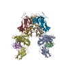
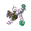
biological assembly 1: chains A, S, Z, B, X, Y, F, E, G, R, D / biological assembly 2: chains P, L ,W, Q, U, V, I, H, J, N, M
-
Components
-
Protein , 9 types, 18 molecules APSLZWBQXUYVFIEHGJ
#1: Protein
U1-A / U1 snRNP A / U1 small nuclear ribonucleoprotein A / U1A / Coordinate model: Cα atoms only
Mass: 31319.533 Da / Num. of mol.: 2 / Source method: isolated from a natural source / Source: (natural) Homo sapiens (human) / Cell line: HELA / References: UniProt: P09012
Mass: 51683.184 Da / Num. of mol.: 2 / Source method: isolated from a natural source / Source: (natural) Homo sapiens (human) / Cell line: HELA / References: UniProt: P08621
#3: Protein
Sm-D3 / Small nuclear ribonucleoprotein Sm D3 / snRNP core protein D3 / Coordinate model: Cα atoms only
Mass: 13940.308 Da / Num. of mol.: 2 / Source method: isolated from a natural source / Source: (natural) Homo sapiens (human) / Cell line: HELA / References: UniProt: P62318
#4: Protein
SmB / Small nuclear ribonucleoprotein polypeptides B and B1 / Coordinate model: Cα atoms only
Mass: 23686.004 Da / Num. of mol.: 2 / Source method: isolated from a natural source / Source: (natural) Homo sapiens (human) / Cell line: HELA / References: UniProt: Q66K91, UniProt: P14678*PLUS
#5: Protein
Sm-D1 / Small nuclear ribonucleoprotein Sm D1 / Sm-D autoantigen / snRNP core protein D1 / Coordinate model: Cα atoms only
Mass: 13310.653 Da / Num. of mol.: 2 / Source method: isolated from a natural source / Source: (natural) Homo sapiens (human) / Cell line: HELA / References: UniProt: P62314
#6: Protein
Sm-D2 / Small nuclear ribonucleoprotein Sm D2 / snRNP core protein D2 / Coordinate model: Cα atoms only
Mass: 13551.928 Da / Num. of mol.: 2 / Source method: isolated from a natural source / Source: (natural) Homo sapiens (human) / Cell line: HELA / References: UniProt: P62316
#7: Protein
Sm-F / snRNP-F / Sm protein F / Small nuclear ribonucleoprotein F / SmF / Coordinate model: Cα atoms only
Mass: 9734.171 Da / Num. of mol.: 2 / Source method: isolated from a natural source / Source: (natural) Homo sapiens (human) / Cell line: HELA / References: UniProt: P62306
#8: Protein
Sm-E / snRNP-E / Sm protein E / Small nuclear ribonucleoprotein E / SmE / Coordinate model: Cα atoms only
Mass: 10817.601 Da / Num. of mol.: 2 / Source method: isolated from a natural source / Source: (natural) Homo sapiens (human) / Cell line: HELA / References: UniProt: P62304
#9: Protein
SmG / Small nuclear ribonucleoprotein polypeptide G / isoform CRA_a / Coordinate model: Cα atoms only
Mass: 8508.084 Da / Num. of mol.: 2 / Source method: isolated from a natural source / Source: (natural) Homo sapiens (human) / Cell line: HELA / References: UniProt: D6W5G6, UniProt: P62308*PLUS
-
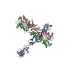
RNA chain / DNA chain , 2 types, 4 molecules RNDM
#10: RNA chain
U1snRNA / U1 spliceosomal RNA / Coordinate model: P atoms only
Mass: 52697.070 Da / Num. of mol.: 2 / Source method: isolated from a natural source / Source: (natural) Homo sapiens (human) / Cell line: HELA
#11: DNA chain
DNA5'-D(*AP*GP*GP*TP*AP*AP*GP*TP*A)-3' / Coordinate model: P atoms only
Mass: 2803.873 Da / Num. of mol.: 2 / Source method: obtained synthetically / Details: DNA oligonucleotide
-
Experimental details
-
Experiment
Experiment
Method: X-RAY DIFFRACTION / Number of used crystals: 1
-
Sample preparation
Crystal
Density Matthews: 2.39 Å3/Da / Density % sol: 48.43 %
Crystal grow
Temperature: 277 K / Method: vapor diffusion, sitting drop / pH: 5.6 Details: 8-10% PEG 4000, 0.1 M Trisodium citrate, 2 mM EDTA, 100 mM NaCl, 1-2% Anapoe X-305, pH 5.6, VAPOR DIFFUSION, SITTING DROP, temperature 277K
In the structure databanks used in Yorodumi, some data are registered as the other names, "COVID-19 virus" and "2019-nCoV". Here are the details of the virus and the list of structure data.
Jan 31, 2019. EMDB accession codes are about to change! (news from PDBe EMDB page)
EMDB accession codes are about to change! (news from PDBe EMDB page)
The allocation of 4 digits for EMDB accession codes will soon come to an end. Whilst these codes will remain in use, new EMDB accession codes will include an additional digit and will expand incrementally as the available range of codes is exhausted. The current 4-digit format prefixed with “EMD-” (i.e. EMD-XXXX) will advance to a 5-digit format (i.e. EMD-XXXXX), and so on. It is currently estimated that the 4-digit codes will be depleted around Spring 2019, at which point the 5-digit format will come into force.
The EM Navigator/Yorodumi systems omit the EMD- prefix.
Related info.:Q: What is EMD? / ID/Accession-code notation in Yorodumi/EM Navigator
Yorodumi is a browser for structure data from EMDB, PDB, SASBDB, etc.
This page is also the successor to EM Navigator detail page, and also detail information page/front-end page for Omokage search.
The word "yorodu" (or yorozu) is an old Japanese word meaning "ten thousand". "mi" (miru) is to see.
Related info.:EMDB / PDB / SASBDB / Comparison of 3 databanks / Yorodumi Search / Aug 31, 2016. New EM Navigator & Yorodumi / Yorodumi Papers / Jmol/JSmol / Function and homology information / Changes in new EM Navigator and Yorodumi
 Movie
Movie Controller


 Open data
 Basic information
 Components
 Keywords
 U1 snRNA / Sm fold / Sm core / RRM /
  Function and homology information
 Authors
 Citation
 Structure visualization
 Downloads & links
 Other downloads
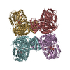


 PDBj
PDBj


 Assembly
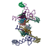

 Sample preparation
 / Beamline: X06SA / Wavelength: 0.999 Å
 Processing