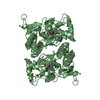
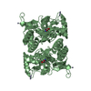
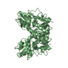
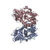
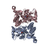
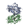
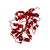
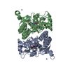
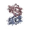
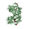
登録情報 データベース : PDB / ID : 3pd9タイトル X-ray structure of the ligand-binding core of GluA2 in complex with (R)-5-HPCA at 2.1 A resolution Glutamate receptor 2 キーワード / / / 機能・相同性 分子機能 ドメイン・相同性 構成要素
/ / / / / / / / / / / / / / / / / / / / / / / / / / / / / / / / / / / / / / / / / / / / / / / / / / / / / / / / / / / / / / / / / / / / / / / / / / / / / / / / / / / / / / / / / / / / / / / / / / / / / / / / / / 生物種 Rattus norvegicus (ドブネズミ)手法 / / / 解像度 : 2.1 Å データ登録者 Frydenvang, K. / Kastrup, J.S. ジャーナル : J. Med. Chem. / 年 : 2010タイトル : Biostructural and pharmacological studies of bicyclic analogues of the 3-isoxazolol glutamate receptor agonist ibotenic acid.
著者 :
Frydenvang, K. / Pickering, D.S. / Greenwood, J.R. / Krogsgaard-Larsen, N. / Brehm, L. / Nielsen, B. / Vogensen, S.B. / Hald, H. / Kastrup, J.S. / Krogsgaard-Larsen, P. / Clausen, R.P. 履歴 登録 2010年10月22日 登録サイト / 処理サイト 改定 1.0 2010年12月29日 Provider / タイプ 改定 1.1 2011年7月13日 Group 改定 1.2 2011年11月9日 Group 改定 1.3 2017年8月23日 Group / カテゴリ 改定 1.4 2018年10月10日 Group / Database references / Structure summaryカテゴリ / diffrn_source / entityItem _citation.journal_abbrev / _citation.journal_id_ISSN ... _citation.journal_abbrev / _citation.journal_id_ISSN / _citation.journal_volume / _citation.page_first / _citation.page_last / _citation.title / _diffrn_source.pdbx_synchrotron_site / _entity.formula_weight 改定 1.5 2023年9月6日 Group Data collection / Database references ... Data collection / Database references / Derived calculations / Refinement description / Structure summary カテゴリ chem_comp_atom / chem_comp_bond ... chem_comp_atom / chem_comp_bond / database_2 / entity / pdbx_initial_refinement_model / struct_ref_seq_dif / struct_site Item _database_2.pdbx_DOI / _database_2.pdbx_database_accession ... _database_2.pdbx_DOI / _database_2.pdbx_database_accession / _entity.formula_weight / _struct_ref_seq_dif.details / _struct_site.pdbx_auth_asym_id / _struct_site.pdbx_auth_comp_id / _struct_site.pdbx_auth_seq_id
すべて表示 表示を減らす
 ムービー
ムービー コントローラー


 データを開く
 基本情報
基本情報 要素
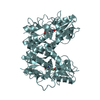
要素 GRIA2
GRIA2  キーワード
キーワード 機能・相同性情報
 データ登録者
 引用
引用 構造の表示
構造の表示 ダウンロードとリンク
 その他のダウンロード
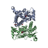


 PDBj
PDBj


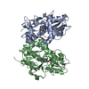
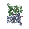
 集合体
集合体

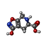


 分子量: 184.149 Da / 分子数: 2 / 由来タイプ: 合成 / 式: C7H8N2O4
 分子量: 96.063 Da / 分子数: 4 / 由来タイプ: 合成 / 式: SO4
 分子量: 92.094 Da / 分子数: 5 / 由来タイプ: 合成 / 式: C3H8O3
 分子量: 35.453 Da / 分子数: 4 / 由来タイプ: 合成 / 式: Cl
 試料調製
試料調製 / ビームライン: X11 / 波長: 0.8126 Å
 解析
解析