 Movie
Movie Controller


+ Open data

- Basic information
| Entry | Database: PDB / ID: 3pd7 | |||||||||
|---|---|---|---|---|---|---|---|---|---|---|
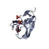
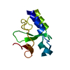
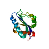
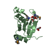
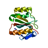
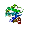
| Title | Crystal Structure of the Sixth BRCT Domain of Human TopBP1 | |||||||||
 Components Components | DNA topoisomerase 2-binding protein 1 | |||||||||
 Keywords Keywords |  CELL CYCLE / BRCT DOMAIN / DNA REPAIR CELL CYCLE / BRCT DOMAIN / DNA REPAIR | |||||||||
| Function / homology |  Function and homology information Function and homology informationbroken chromosome clustering / BRCA1-B complex / phosphorylation-dependent protein binding / chromatin-protein adaptor activity / homologous recombination / protein localization to site of double-strand break / DNA replication checkpoint signaling / double-strand break repair via classical nonhomologous end joining / mitotic DNA replication checkpoint signaling / double-strand break repair via alternative nonhomologous end joining ...broken chromosome clustering / BRCA1-B complex / phosphorylation-dependent protein binding / chromatin-protein adaptor activity / homologous recombination / protein localization to site of double-strand break / DNA replication checkpoint signaling / double-strand break repair via classical nonhomologous end joining / mitotic DNA replication checkpoint signaling / double-strand break repair via alternative nonhomologous end joining / HDR through Single Strand Annealing (SSA) / Impaired BRCA2 binding to RAD51 / DNA metabolic process / response to ionizing radiation / mitotic G2 DNA damage checkpoint signaling / site of DNA damage / Presynaptic phase of homologous DNA pairing and strand exchange / chromosome organization / DNA replication initiation / protein serine/threonine kinase activator activity / DNA damage checkpoint signaling / condensed nuclear chromosome / male germ cell nucleus / double-strand break repair via homologous recombination / G2/M DNA damage checkpoint / PML body / spindle pole / actin cytoskeleton / site of double-strand break / chromosome / Processing of DNA double-strand break ends / Regulation of TP53 Activity through Phosphorylation / nuclear body / DNA repair / centrosome / DNA damage response / DNA binding / nucleoplasm / identical protein binding / nucleus / plasma membrane / cytoplasmSimilarity search - Function | |||||||||
| Biological species |  Homo sapiens (human) Homo sapiens (human) | |||||||||
| Method | X-RAY DIFFRACTION / SYNCHROTRON / MOLECULAR REPLACEMENT / Resolution: 1.26 Å | |||||||||
 Authors Authors | Lee, J. / Xu, C. / Cui, G. / Thompson, J.R. / Mer, G. | |||||||||
 Citation Citation | Journal: To be Published Title: Crystal Structure of the Sixth BRCT Domain of Human TopBP1 Authors: Lee, J. / Xu, C. / Cui, G. / Thompson, J.R. / Mer, G. | |||||||||
| History |
|
- Structure visualization
| Structure viewer | Molecule: MolmilJmol/JSmol |
|---|
- Downloads & links
-Download
| PDBx/mmCIF format | 3pd7.cif.gz | 146.9 KB | Display | PDBx/mmCIF format |
|---|---|---|---|---|
| PDB format | pdb3pd7.ent.gz | 119.2 KB | Display | PDB format |
| PDBx/mmJSON format | 3pd7.json.gz | Tree view | PDBx/mmJSON format | |
| Others |  Other downloads Other downloads |
-Validation report
| Arichive directory | https://data.pdbj.org/pub/pdb/validation_reports/pd/3pd7ftp://data.pdbj.org/pub/pdb/validation_reports/pd/3pd7 | HTTPS FTP |
|---|
-Related structure data
| Similar structure data |
|---|


-Links
 PDBj
PDBj


- Assembly
| Deposited unit | 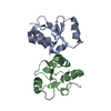
| |||||||||||||||||||||
|---|---|---|---|---|---|---|---|---|---|---|---|---|---|---|---|---|---|---|---|---|---|---|
| 1 | 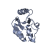
| |||||||||||||||||||||
| 2 | 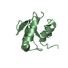
| |||||||||||||||||||||
| Unit cell |
| |||||||||||||||||||||
| Components on special symmetry positions |
|
-Components
| #1: Protein | Mass: 12216.718 Da / Num. of mol.: 2 / Fragment: Sixth BRCT domain, UNP residues 893-994 Source method: isolated from a genetically manipulated source Source: (gene. exp.) Homo sapiens (human) / Gene: TOPBP1, KIAA0259 / Plasmid: pTEV / Production host:  Escherichia coli (E. coli) / Strain (production host): BL21(DE3) / References: UniProt: Q92547 Escherichia coli (E. coli) / Strain (production host): BL21(DE3) / References: UniProt: Q92547#2: Water | ChemComp-HOH / | Water Mass: 18.015 Da / Num. of mol.: 357 / Source method: isolated from a natural source / Formula: H2O Mass: 18.015 Da / Num. of mol.: 357 / Source method: isolated from a natural source / Formula: H2O |
|---|
-Experimental details
-Experiment
| Experiment | Method: X-RAY DIFFRACTION / Number of used crystals: 1 |
|---|
- Sample preparation
| Crystal | Density Matthews: 2.03 Å3/Da / Density % sol: 39.29 % |
|---|---|
| Crystal grow | Temperature: 295 K / Method: vapor diffusion, hanging drop / pH: 6.6 Details: 20% PEG 4000, 0.2 M sodium acetate trihydrate, 0.1 M Tris-HCl, pH 6.6, VAPOR DIFFUSION, HANGING DROP, temperature 295K |
-Data collection
| Diffraction | Mean temperature: 100 K |
|---|---|
| Diffraction source | Source: SYNCHROTRON / Site: APS  / Beamline: 19-BM / Wavelength: 1.0079 Å / Beamline: 19-BM / Wavelength: 1.0079 Å |
| Detector | Type: ADSC QUANTUM 210r / Detector: CCD / Date: Feb 10, 2006 |
| Radiation | Protocol: SINGLE WAVELENGTH / Monochromatic (M) / Laue (L): M / Scattering type: x-ray |
| Radiation wavelength | Wavelength: 1.0079 Å / Relative weight: 1 |
| Reflection | Resolution: 1.26→50 Å / Num. obs: 51641 / % possible obs: 100 % / Redundancy: 9.2 % / Rmerge(I) obs: 0.039 / Net I/σ(I): 53.6 |
| Reflection shell | Resolution: 1.26→1.31 Å / Redundancy: 8.7 % / Rmerge(I) obs: 0.242 / Mean I/σ(I) obs: 8.4 / Num. unique all: 9747 / % possible all: 99.8 |
- Processing
| Software |
| ||||||||||||||||||||||||||||||||||||||||||
|---|---|---|---|---|---|---|---|---|---|---|---|---|---|---|---|---|---|---|---|---|---|---|---|---|---|---|---|---|---|---|---|---|---|---|---|---|---|---|---|---|---|---|---|
| Refinement | Method to determine structure: MOLECULAR REPLACEMENT / Resolution: 1.26→25.322 Å / SU ML: 0.15 / σ(F): 1.37 / Stereochemistry target values: ML
| ||||||||||||||||||||||||||||||||||||||||||
| Solvent computation | Shrinkage radii: 0.9 Å / VDW probe radii: 1.11 Å / Solvent model: FLAT BULK SOLVENT MODEL / Bsol: 45.479 Å2 / ksol: 0.376 e/Å3 | ||||||||||||||||||||||||||||||||||||||||||
| Displacement parameters |
| ||||||||||||||||||||||||||||||||||||||||||
| Refinement step | Cycle: LAST / Resolution: 1.26→25.322 Å
| ||||||||||||||||||||||||||||||||||||||||||
| Refine LS restraints |
| ||||||||||||||||||||||||||||||||||||||||||
| LS refinement shell |
|
