 Movie
Movie Controller


+ Open data

- Basic information
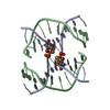
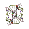
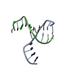
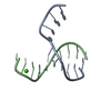
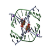
| Entry | Database: PDB / ID: 3pbx | ||||||
|---|---|---|---|---|---|---|---|
| Title | Strontium bound to the sequence d(CCGGCGCCGG) | ||||||
 Components Components | DNA | ||||||
 Keywords Keywords | DNA / Holliday junction (higher order DNA structure) / Intermediate formed in Homologous recombination | ||||||
| Function / homology | STRONTIUM ION / DNA Function and homology information Function and homology information | ||||||
| Method | X-RAY DIFFRACTION / MOLECULAR REPLACEMENT / Resolution: 1.879 Å | ||||||
 Authors Authors | Naseer, A. / Cardin, C.J. | ||||||
 Citation Citation | Journal: To be Published Title: Strontium bound to the sequence d(CCGGCGCCGG) Authors: Naseer, A. / Cardin, C.J. | ||||||
| History |
|
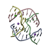
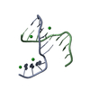
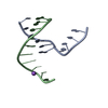
- Structure visualization
| Structure viewer | Molecule: MolmilJmol/JSmol |
|---|
- Downloads & links
-Download
| PDBx/mmCIF format | 3pbx.cif.gz | 22.2 KB | Display | PDBx/mmCIF format |
|---|---|---|---|---|
| PDB format | pdb3pbx.ent.gz | 15 KB | Display | PDB format |
| PDBx/mmJSON format | 3pbx.json.gz | Tree view | PDBx/mmJSON format | |
| Others |  Other downloads Other downloads |
-Validation report
| Arichive directory | https://data.pdbj.org/pub/pdb/validation_reports/pb/3pbxftp://data.pdbj.org/pub/pdb/validation_reports/pb/3pbx | HTTPS FTP |
|---|
-Related structure data
| Related structure data |  3gojS S: Starting model for refinement |
|---|---|
| Similar structure data |


-Links
 PDBj
PDBj


- Assembly
| Deposited unit | 
| ||||||||
|---|---|---|---|---|---|---|---|---|---|
| 1 | 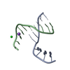
| ||||||||
| Unit cell |
| ||||||||
| Components on special symmetry positions |
|
-Components
| #1: DNA chain | Mass: 3046.980 Da / Num. of mol.: 2 / Source method: obtained synthetically / Details: Chemically synthesized #2: Chemical | ChemComp-SR / | Strontium  Mass: 87.620 Da / Num. of mol.: 1 / Source method: obtained synthetically / Formula: Sr Mass: 87.620 Da / Num. of mol.: 1 / Source method: obtained synthetically / Formula: Sr#3: Chemical | ChemComp-NA / |   Mass: 22.990 Da / Num. of mol.: 1 / Source method: obtained synthetically / Formula: Na Mass: 22.990 Da / Num. of mol.: 1 / Source method: obtained synthetically / Formula: Na#4: Water | ChemComp-HOH / | Water Mass: 18.015 Da / Num. of mol.: 57 / Source method: isolated from a natural source / Formula: H2O Mass: 18.015 Da / Num. of mol.: 57 / Source method: isolated from a natural source / Formula: H2O |
|---|
-Experimental details
-Experiment
| Experiment | Method: X-RAY DIFFRACTION / Number of used crystals: 1 |
|---|
- Sample preparation
| Crystal | Density Matthews: 2.32 Å3/Da / Density % sol: 46.9 % |
|---|---|
| Crystal grow | Temperature: 290 K / Method: vapor diffusion, sitting drop / pH: 7 Details: SrCl2, MPD, NaCacodylate , pH 7.0, VAPOR DIFFUSION, SITTING DROP, temperature 290K |
-Data collection
| Diffraction | Mean temperature: 100 K |
|---|---|
| Diffraction source | Source: SEALED TUBE / Type: OXFORD DIFFRACTION ENHANCE ULTRA / Wavelength: 1.5418 Å |
| Detector | Type: OXFORD SAPPHIRE CCD / Detector: CCD / Date: Feb 10, 2007 / Details: multi-graded optics |
| Radiation | Protocol: SINGLE WAVELENGTH / Monochromatic (M) / Laue (L): M / Scattering type: x-ray |
| Radiation wavelength | Wavelength: 1.5418 Å / Relative weight: 1 |
| Reflection | Resolution: 1.879→14.866 Å / Num. all: 4705 / Num. obs: 3706 / % possible obs: 78.55 % / Observed criterion σ(F): 1 / Observed criterion σ(I): 1 / Redundancy: 2.1 % / Biso Wilson estimate: 27.446 Å2 / Rmerge(I) obs: 0.045 |
| Reflection shell | Resolution: 1.879→1.927 Å |
- Processing
| Software |
| |||||||||||||||||||||||||||||||||||||||||||||||||||||||||||||||||
|---|---|---|---|---|---|---|---|---|---|---|---|---|---|---|---|---|---|---|---|---|---|---|---|---|---|---|---|---|---|---|---|---|---|---|---|---|---|---|---|---|---|---|---|---|---|---|---|---|---|---|---|---|---|---|---|---|---|---|---|---|---|---|---|---|---|---|
| Refinement | Method to determine structure: MOLECULAR REPLACEMENT Starting model: 3GOJ Resolution: 1.879→14.86 Å / Cor.coef. Fo:Fc: 0.953 / Cor.coef. Fo:Fc free: 0.872 / SU B: 5.747 / SU ML: 0.153 / Cross valid method: THROUGHOUT / ESU R: 0.273 / ESU R Free: 0.245 / Stereochemistry target values: MAXIMUM LIKELIHOOD / Details: HYDROGENS HAVE BEEN ADDED IN THE RIDING POSITIONS
| |||||||||||||||||||||||||||||||||||||||||||||||||||||||||||||||||
| Solvent computation | Ion probe radii: 0.8 Å / Shrinkage radii: 0.8 Å / VDW probe radii: 1.2 Å / Solvent model: MASK | |||||||||||||||||||||||||||||||||||||||||||||||||||||||||||||||||
| Displacement parameters | Biso mean: 27.446 Å2
| |||||||||||||||||||||||||||||||||||||||||||||||||||||||||||||||||
| Refinement step | Cycle: LAST / Resolution: 1.879→14.86 Å
| |||||||||||||||||||||||||||||||||||||||||||||||||||||||||||||||||
| Refine LS restraints |
| |||||||||||||||||||||||||||||||||||||||||||||||||||||||||||||||||
| LS refinement shell | Resolution: 1.879→1.927 Å / Total num. of bins used: 20
|
