 Movie
Movie Controller

[English] 日本語
 Yorodumi
- PDB-3pb0: Characterisation of the first monomeric dihydrodipicolinate synth... -
+ Open data

- Basic information
| Entry | Database: PDB / ID: 3pb0 | ||||||
|---|---|---|---|---|---|---|---|
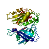
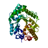
| Title | Characterisation of the first monomeric dihydrodipicolinate synthase variant reveals evolutionary insights | ||||||
 Components Components | Dihydrodipicolinate synthase | ||||||
 Keywords Keywords | LYASE / dihydrodipicolinate synthase | ||||||
| Function / homology |  Function and homology information4-hydroxy-2-oxoglutarate aldolase activity / 4-hydroxy-tetrahydrodipicolinate synthase / 4-hydroxy-tetrahydrodipicolinate synthase activity / glyoxylate catabolic process / diaminopimelate biosynthetic process / lysine biosynthetic process via diaminopimelate / cytosol Function and homology information4-hydroxy-2-oxoglutarate aldolase activity / 4-hydroxy-tetrahydrodipicolinate synthase / 4-hydroxy-tetrahydrodipicolinate synthase activity / glyoxylate catabolic process / diaminopimelate biosynthetic process / lysine biosynthetic process via diaminopimelate / cytosolSimilarity search - Function | ||||||
| Biological species |   thermotoga maritima (bacteria) thermotoga maritima (bacteria) | ||||||
| Method | X-RAY DIFFRACTION / SYNCHROTRON / MOLECULAR REPLACEMENT / Resolution: 2 Å | ||||||
 Authors Authors | Pearce, F.G. / Dobson, R.C.J. / Jameson, G.B. | ||||||
 Citation Citation | Journal: Biochim.Biophys.Acta / Year: 2011 Title: Characterization of monomeric dihydrodipicolinate synthase variant reveals the importance of substrate binding in optimizing oligomerization. Authors: Pearce, F.G. / Dobson, R.C. / Jameson, G.B. / Perugini, M.A. / Gerrard, J.A. | ||||||
| History |
|
- Structure visualization
| Structure viewer | Molecule: MolmilJmol/JSmol |
|---|
- Downloads & links
-Download
| PDBx/mmCIF format | 3pb0.cif.gz | 259.5 KB | Display | PDBx/mmCIF format |
|---|---|---|---|---|
| PDB format | pdb3pb0.ent.gz | 208.1 KB | Display | PDB format |
| PDBx/mmJSON format | 3pb0.json.gz | Tree view | PDBx/mmJSON format | |
| Others |  Other downloads Other downloads |
-Validation report
| Arichive directory | https://data.pdbj.org/pub/pdb/validation_reports/pb/3pb0ftp://data.pdbj.org/pub/pdb/validation_reports/pb/3pb0 | HTTPS FTP |
|---|
-Related structure data


-Links
 PDBj
PDBj- Assembly
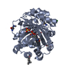
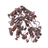
| Deposited unit | 
| ||||||||
|---|---|---|---|---|---|---|---|---|---|
| 1 | 
| ||||||||
| 2 | 
| ||||||||
| 3 | 
| ||||||||
| 4 | 
| ||||||||
| Unit cell |
|
-Components
| #1: Protein | / DHDPS Mass: 32927.801 Da / Num. of mol.: 4 / Mutation: D166A, I167A, D168A Source method: isolated from a genetically manipulated source Source: (gene. exp.) thermotoga maritima (bacteria) / Production host: Escherichia coli (E. coli) / References: UniProt: Q9X1K9, dihydrodipicolinate synthase#2: Chemical | Sulfate  Mass: 96.063 Da / Num. of mol.: 3 / Source method: obtained synthetically / Formula: SO4 Mass: 96.063 Da / Num. of mol.: 3 / Source method: obtained synthetically / Formula: SO4#3: Water | ChemComp-HOH / | Water Mass: 18.015 Da / Num. of mol.: 1064 / Source method: isolated from a natural source / Formula: H2O Mass: 18.015 Da / Num. of mol.: 1064 / Source method: isolated from a natural source / Formula: H2O |
|---|
-Experimental details
-Experiment
| Experiment | Method: X-RAY DIFFRACTION |
|---|
- Sample preparation
| Crystal | Density Matthews: 2.6 Å3/Da / Density % sol: 52.64 % |
|---|---|
| Crystal grow | Temperature: 293 K / Method: vapor diffusion, sitting drop / pH: 4.5 Details: 200mM Lithium sulphate, 50% v/v PEG 400, 100mM sodium-citrate buffer, pH 4.5, VAPOR DIFFUSION, SITTING DROP, temperature 293K |
-Data collection
| Diffraction | Mean temperature: 110 K | |||||||||||||||
|---|---|---|---|---|---|---|---|---|---|---|---|---|---|---|---|---|
| Diffraction source | Source: SYNCHROTRON / Site: Australian Synchrotron  / Beamline: MX2 / Wavelength: 0.9536 Å / Beamline: MX2 / Wavelength: 0.9536 Å | |||||||||||||||
| Detector | Type: ADSC QUANTUM 315r / Detector: CCD / Date: Jan 1, 2006 | |||||||||||||||
| Radiation | Monochromator: YALE MIRRORS / Protocol: SINGLE WAVELENGTH / Monochromatic (M) / Laue (L): M / Scattering type: x-ray | |||||||||||||||
| Radiation wavelength | Wavelength: 0.9536 Å / Relative weight: 1 | |||||||||||||||
| Reflection twin |
| |||||||||||||||
| Reflection | Resolution: 2→74.16 Å / Num. all: 90641 / Num. obs: 82574 / % possible obs: 91.1 % / Observed criterion σ(F): 1 / Observed criterion σ(I): 1 | |||||||||||||||
| Reflection shell | Resolution: 2→2.11 Å / % possible all: 79.1 |
- Processing
| Software |
| ||||||||||||||||||||||||||||||||||||||||||||||||||||||||||||||||||||||||||||||||||||||||||||||||||||||||||||||||||||||||||||||||||||||||||||||||||||||||||||||||||||||||||
|---|---|---|---|---|---|---|---|---|---|---|---|---|---|---|---|---|---|---|---|---|---|---|---|---|---|---|---|---|---|---|---|---|---|---|---|---|---|---|---|---|---|---|---|---|---|---|---|---|---|---|---|---|---|---|---|---|---|---|---|---|---|---|---|---|---|---|---|---|---|---|---|---|---|---|---|---|---|---|---|---|---|---|---|---|---|---|---|---|---|---|---|---|---|---|---|---|---|---|---|---|---|---|---|---|---|---|---|---|---|---|---|---|---|---|---|---|---|---|---|---|---|---|---|---|---|---|---|---|---|---|---|---|---|---|---|---|---|---|---|---|---|---|---|---|---|---|---|---|---|---|---|---|---|---|---|---|---|---|---|---|---|---|---|---|---|---|---|---|---|---|---|
| Refinement | Method to determine structure: MOLECULAR REPLACEMENT / Resolution: 2→37.21 Å / Cor.coef. Fo:Fc: 0.961 / Cor.coef. Fo:Fc free: 0.93 / SU B: 4.469 / SU ML: 0.115 / Cross valid method: THROUGHOUT / σ(F): 1 / ESU R Free: 0.032 / Stereochemistry target values: MAXIMUM LIKELIHOOD / Details: HYDROGENS HAVE BEEN ADDED IN THE RIDING POSITIONS
| ||||||||||||||||||||||||||||||||||||||||||||||||||||||||||||||||||||||||||||||||||||||||||||||||||||||||||||||||||||||||||||||||||||||||||||||||||||||||||||||||||||||||||
| Solvent computation | Ion probe radii: 0.8 Å / Shrinkage radii: 0.8 Å / VDW probe radii: 1.4 Å / Solvent model: MASK | ||||||||||||||||||||||||||||||||||||||||||||||||||||||||||||||||||||||||||||||||||||||||||||||||||||||||||||||||||||||||||||||||||||||||||||||||||||||||||||||||||||||||||
| Displacement parameters | Biso mean: 26.888 Å2
| ||||||||||||||||||||||||||||||||||||||||||||||||||||||||||||||||||||||||||||||||||||||||||||||||||||||||||||||||||||||||||||||||||||||||||||||||||||||||||||||||||||||||||
| Refinement step | Cycle: LAST / Resolution: 2→37.21 Å
| ||||||||||||||||||||||||||||||||||||||||||||||||||||||||||||||||||||||||||||||||||||||||||||||||||||||||||||||||||||||||||||||||||||||||||||||||||||||||||||||||||||||||||
| Refine LS restraints |
| ||||||||||||||||||||||||||||||||||||||||||||||||||||||||||||||||||||||||||||||||||||||||||||||||||||||||||||||||||||||||||||||||||||||||||||||||||||||||||||||||||||||||||
| LS refinement shell | Resolution: 1.999→2.051 Å / Total num. of bins used: 20
|
