Mass: 18.015 Da / Num. of mol.: 420 / Source method: isolated from a natural source / Formula: H2O
-
Details
Nonpolymer details
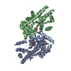
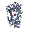
THE STARTING INACTIVATOR MOLECULE (S)-4-AMINO-4,5-DYHIDRO-2-FURANCARBOXYLIC ACID (S-ADFA) UNDERGOES ...THE STARTING INACTIVATOR MOLECULE (S)-4-AMINO-4,5-DYHIDRO-2-FURANCARBOXYLIC ACID (S-ADFA) UNDERGOES A CHEMICAL TRANSFORMATION BY IRREVERSIBLY BINDING TO THE ACTIVE SITE RESIDUE K246 OF ASPC. THE LIGAND PJ7 REPRESENTS FINAL PRODUCT
-
Experimental details
-
Experiment
Experiment
Method: X-RAY DIFFRACTION / Number of used crystals: 1
-
Sample preparation
Crystal
Density Matthews: 2.99 Å3/Da / Density % sol: 58.85 %
Crystal grow
Temperature: 297 K / Method: vapor diffusion / pH: 7.5 Details: Ammonium Sulfate, pH 7.5, VAPOR DIFFUSION, temperature 297K
Resolution: 1.7→77.15 Å / Cor.coef. Fo:Fc: 0.972 / Cor.coef. Fo:Fc free: 0.961 / SU B: 3.485 / SU ML: 0.053 / Cross valid method: THROUGHOUT / σ(F): 2 / ESU R Free: 0.087 / Stereochemistry target values: MAXIMUM LIKELIHOOD / Details: HYDROGENS HAVE BEEN ADDED IN THE RIDING POSITIONS
Rfactor
Num. reflection
% reflection
Selection details
Rfree
0.18519
2851
5.1 %
RANDOM
Rwork
0.14827
-
-
-
obs
0.15012
53395
97.83 %
-
all
-
67102
-
-
Solvent computation
Ion probe radii: 0.8 Å / Shrinkage radii: 0.8 Å / VDW probe radii: 1.4 Å / Solvent model: MASK
Displacement parameters
Biso mean: 23.043 Å2
Baniso -1
Baniso -2
Baniso -3
1-
-0.59 Å2
0 Å2
0 Å2
2-
-
0.55 Å2
0 Å2
3-
-
-
0.04 Å2
Refinement step
Cycle: LAST / Resolution: 1.7→77.15 Å
Protein
Nucleic acid
Ligand
Solvent
Total
Num. atoms
3069
0
110
420
3599
Refine LS restraints
Refine-ID
Type
Dev ideal
Dev ideal target
Number
X-RAY DIFFRACTION
r_bond_refined_d
0.01
0.022
3580
X-RAY DIFFRACTION
r_angle_refined_deg
1.269
1.989
4898
X-RAY DIFFRACTION
r_dihedral_angle_1_deg
5.6
5
482
X-RAY DIFFRACTION
r_dihedral_angle_2_deg
34.14
24.253
174
X-RAY DIFFRACTION
r_dihedral_angle_3_deg
13.484
15
612
X-RAY DIFFRACTION
r_dihedral_angle_4_deg
16.314
15
28
X-RAY DIFFRACTION
r_chiral_restr
0.085
0.2
536
X-RAY DIFFRACTION
r_gen_planes_refined
0.005
0.02
2755
X-RAY DIFFRACTION
r_nbd_refined
0.22
0.2
1852
X-RAY DIFFRACTION
r_nbtor_refined
0.307
0.2
2444
X-RAY DIFFRACTION
r_xyhbond_nbd_refined
0.128
0.2
380
X-RAY DIFFRACTION
r_symmetry_vdw_refined
0.194
0.2
140
X-RAY DIFFRACTION
r_symmetry_hbond_refined
0.121
0.2
43
X-RAY DIFFRACTION
r_mcbond_it
0.974
1.5
2186
X-RAY DIFFRACTION
r_mcangle_it
1.543
2
3474
X-RAY DIFFRACTION
r_scbond_it
2.445
3
1543
X-RAY DIFFRACTION
r_scangle_it
3.552
4.5
1381
LS refinement shell
Resolution: 1.702→1.746 Å / Total num. of bins used: 20
Rfactor
Num. reflection
% reflection
Rfree
0.279
206
-
Rwork
0.195
3775
-
obs
-
-
94.43 %
+
About Yorodumi
-
News
-
Feb 9, 2022. New format data for meta-information of EMDB entries
New format data for meta-information of EMDB entries
Version 3 of the EMDB header file is now the official format.
The previous official version 1.9 will be removed from the archive.
In the structure databanks used in Yorodumi, some data are registered as the other names, "COVID-19 virus" and "2019-nCoV". Here are the details of the virus and the list of structure data.
Jan 31, 2019. EMDB accession codes are about to change! (news from PDBe EMDB page)
EMDB accession codes are about to change! (news from PDBe EMDB page)
The allocation of 4 digits for EMDB accession codes will soon come to an end. Whilst these codes will remain in use, new EMDB accession codes will include an additional digit and will expand incrementally as the available range of codes is exhausted. The current 4-digit format prefixed with “EMD-” (i.e. EMD-XXXX) will advance to a 5-digit format (i.e. EMD-XXXXX), and so on. It is currently estimated that the 4-digit codes will be depleted around Spring 2019, at which point the 5-digit format will come into force.
The EM Navigator/Yorodumi systems omit the EMD- prefix.
Related info.:Q: What is EMD? / ID/Accession-code notation in Yorodumi/EM Navigator
Yorodumi is a browser for structure data from EMDB, PDB, SASBDB, etc.
This page is also the successor to EM Navigator detail page, and also detail information page/front-end page for Omokage search.
The word "yorodu" (or yorozu) is an old Japanese word meaning "ten thousand". "mi" (miru) is to see.
Related info.:EMDB / PDB / SASBDB / Comparison of 3 databanks / Yorodumi Search / Aug 31, 2016. New EM Navigator & Yorodumi / Yorodumi Papers / Jmol/JSmol / Function and homology information / Changes in new EM Navigator and Yorodumi
 Movie
Movie Controller

 Yorodumi
 Open data
 Basic information
 Components
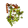
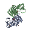
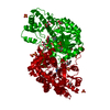
 Aspartate transaminase
  Keywords
 Function and homology information
 Authors
 Citation
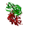
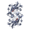
 Structure visualization
 Downloads & links
 Other downloads


 PDBj
PDBj Assembly


 Mass: 127.098 Da / Num. of mol.: 1 / Source method: obtained synthetically / Formula: C5H5NO3
 Mass: 248.173 Da / Num. of mol.: 1 / Source method: obtained synthetically / Formula: C8H13N2O5P
 Mass: 92.094 Da / Num. of mol.: 11 / Source method: obtained synthetically / Formula: C3H8O3
 Mass: 96.063 Da / Num. of mol.: 4 / Source method: obtained synthetically / Formula: SO4
 Sample preparation
 / Beamline: 14-BM-C / Wavelength: 0.97 Å
 Processing