 Movie
Movie Controller

[English] 日本語
 Yorodumi
- PDB-3pa7: Crystal structure of FKBP from plasmodium vivax in complex with t... -
+ Open data

- Basic information
| Entry | Database: PDB / ID: 3pa7 | ||||||
|---|---|---|---|---|---|---|---|
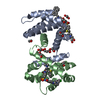
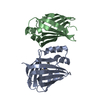
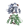
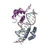
| Title | Crystal structure of FKBP from plasmodium vivax in complex with tetrapeptide ALPF | ||||||
 Components Components |
| ||||||
 Keywords Keywords |  ISOMERASE / Plasmodium vivax / FKBP35 / PPIase / FK506 binding protein ISOMERASE / Plasmodium vivax / FKBP35 / PPIase / FK506 binding protein | ||||||
| Function / homology |  Function and homology information Function and homology information | ||||||
| Biological species |  Plasmodium vivax (malaria parasite P. vivax) Plasmodium vivax (malaria parasite P. vivax) | ||||||
| Method | X-RAY DIFFRACTION / MOLECULAR REPLACEMENT / Resolution: 2.61 Å | ||||||
 Authors Authors | Balakrishna, A.M. / Alag, R. / Yoon, H.S. | ||||||
 Citation Citation | Journal: EUKARYOTIC CELL / Year: 2013 Title: Structural insights into substrate binding by PvFKBP35, a peptidylprolyl cis-trans isomerase from the human malarial parasite Plasmodium vivax Authors: Alag, R. / Balakrishna, A.M. / Rajan, S. / Qureshi, I.A. / Shin, J. / Lescar, J. / Gruber, G. / Yoon, H.S. #1: Journal: Protein Sci. / Year: 2010Title: NMR and crystallographic structures of the FK506 binding domain of human malarial parasite Plasmodium vivax FKBP35 Authors: Alag, R. / Qureshi, I.A. / Bharatham, N. / Shin, J. / Lescar, J. / Yoon, H.S. | ||||||
| History |
|
- Structure visualization
| Structure viewer | Molecule: MolmilJmol/JSmol |
|---|
- Downloads & links
-Download
| PDBx/mmCIF format | 3pa7.cif.gz | 98.3 KB | Display | PDBx/mmCIF format |
|---|---|---|---|---|
| PDB format | pdb3pa7.ent.gz | 73.7 KB | Display | PDB format |
| PDBx/mmJSON format | 3pa7.json.gz | Tree view | PDBx/mmJSON format | |
| Others |  Other downloads Other downloads |
-Validation report
| Arichive directory | https://data.pdbj.org/pub/pdb/validation_reports/pa/3pa7ftp://data.pdbj.org/pub/pdb/validation_reports/pa/3pa7 | HTTPS FTP |
|---|
-Related structure data
| Related structure data | 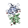 3ni6C 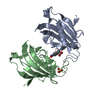 4itzC 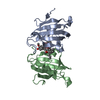 3ihzS S: Starting model for refinement C: citing same article ( |
|---|---|
| Similar structure data |


-Links
 PDBj
PDBj


- Assembly
| Deposited unit | 
| ||||||||||||||||||
|---|---|---|---|---|---|---|---|---|---|---|---|---|---|---|---|---|---|---|---|
| 1 |
| ||||||||||||||||||
| Unit cell |
| ||||||||||||||||||
| Noncrystallographic symmetry (NCS) | NCS domain:
NCS domain segments: Component-ID: 1 / Ens-ID: 1 / Beg auth comp-ID: THR / Beg label comp-ID: THR / End auth comp-ID: ARG / End label comp-ID: ARG / Refine code: 1 / Auth seq-ID: 5 - 125 / Label seq-ID: 5 - 125
|
-Components
| #1: Protein | Mass: 13971.687 Da / Num. of mol.: 2 / Fragment: residues 1-126 Source method: isolated from a genetically manipulated source Source: (gene. exp.) Plasmodium vivax (malaria parasite P. vivax)Strain: SALVADOR I / Gene: PVX_101260 / Plasmid: pSUMO / Production host:  Escherichia coli (E. coli) / Strain (production host): BL21(DE3) / References: UniProt: A5K8X6, peptidylprolyl isomerase Escherichia coli (E. coli) / Strain (production host): BL21(DE3) / References: UniProt: A5K8X6, peptidylprolyl isomerase#2: Protein/peptide | | Mass: 446.539 Da / Num. of mol.: 1 / Source method: obtained synthetically / Details: synthetic peptide #3: Water | ChemComp-HOH / | Water Mass: 18.015 Da / Num. of mol.: 150 / Source method: isolated from a natural source / Formula: H2O Mass: 18.015 Da / Num. of mol.: 150 / Source method: isolated from a natural source / Formula: H2O |
|---|
-Experimental details
-Experiment
| Experiment | Method: X-RAY DIFFRACTION / Number of used crystals: 1 |
|---|
- Sample preparation
| Crystal | Density Matthews: 1.98 Å3/Da / Density % sol: 37.99 % |
|---|---|
| Crystal grow | Temperature: 291 K / Method: vapor diffusion, sitting drop / pH: 9 Details: 100mM BICINE, 2.4M Ammonium sulfate, pH 9.0, VAPOR DIFFUSION, SITTING DROP, temperature 291K |
-Data collection
| Diffraction | Mean temperature: 113 K |
|---|---|
| Diffraction source | Source: ROTATING ANODE / Type: RIGAKU MICROMAX-007 HF / Wavelength: 1.54178 Å |
| Detector | Type: RIGAKU RAXIS IV++ / Detector: IMAGE PLATE / Date: Jul 30, 2010 / Details: mirrors |
| Radiation | Protocol: SINGLE WAVELENGTH / Monochromatic (M) / Laue (L): M / Scattering type: x-ray |
| Radiation wavelength | Wavelength: 1.54178 Å / Relative weight: 1 |
| Reflection | Resolution: 2.61→30 Å / Num. obs: 6670 / % possible obs: 96.8 % / Observed criterion σ(F): 2 / Observed criterion σ(I): 2 / Redundancy: 2.7 % / Biso Wilson estimate: 49.2 Å2 / Rmerge(I) obs: 0.028 / Net I/σ(I): 22.03 |
| Reflection shell | Resolution: 2.61→2.74 Å / Redundancy: 2.7 % / Rmerge(I) obs: 0.028 / Mean I/σ(I) obs: 22.03 / Num. unique all: 6660 / % possible all: 89.9 |
- Processing
| Software |
| |||||||||||||||||||||||||||||||||||||||||||||||||||||||||||||||||||||||||||
|---|---|---|---|---|---|---|---|---|---|---|---|---|---|---|---|---|---|---|---|---|---|---|---|---|---|---|---|---|---|---|---|---|---|---|---|---|---|---|---|---|---|---|---|---|---|---|---|---|---|---|---|---|---|---|---|---|---|---|---|---|---|---|---|---|---|---|---|---|---|---|---|---|---|---|---|---|
| Refinement | Method to determine structure: MOLECULAR REPLACEMENT Starting model: 3IHZ Resolution: 2.61→24.12 Å / Cor.coef. Fo:Fc: 0.909 / Cor.coef. Fo:Fc free: 0.879 / Occupancy max: 1 / Occupancy min: 1 / SU B: 35.665 / SU ML: 0.345 / Cross valid method: THROUGHOUT / σ(F): 0 / ESU R Free: 0.483 / Stereochemistry target values: MAXIMUM LIKELIHOOD Details: HYDROGENS HAVE BEEN ADDED IN THE RIDING POSITIONS U VALUES: RESIDUAL ONLY
| |||||||||||||||||||||||||||||||||||||||||||||||||||||||||||||||||||||||||||
| Solvent computation | Ion probe radii: 0.8 Å / Shrinkage radii: 0.8 Å / VDW probe radii: 1.4 Å / Solvent model: MASK | |||||||||||||||||||||||||||||||||||||||||||||||||||||||||||||||||||||||||||
| Displacement parameters | Biso max: 99.51 Å2 / Biso mean: 41.4839 Å2 / Biso min: 2 Å2
| |||||||||||||||||||||||||||||||||||||||||||||||||||||||||||||||||||||||||||
| Refinement step | Cycle: LAST / Resolution: 2.61→24.12 Å
| |||||||||||||||||||||||||||||||||||||||||||||||||||||||||||||||||||||||||||
| Refine LS restraints |
| |||||||||||||||||||||||||||||||||||||||||||||||||||||||||||||||||||||||||||
| Refine LS restraints NCS | Dom-ID: 1 / Auth asym-ID: A / Ens-ID: 1 / Number: 934 / Refine-ID: X-RAY DIFFRACTION
| |||||||||||||||||||||||||||||||||||||||||||||||||||||||||||||||||||||||||||
| LS refinement shell | Resolution: 2.607→2.674 Å / Total num. of bins used: 20
| |||||||||||||||||||||||||||||||||||||||||||||||||||||||||||||||||||||||||||
| Refinement TLS params. | Method: refined / Refine-ID: X-RAY DIFFRACTION
| |||||||||||||||||||||||||||||||||||||||||||||||||||||||||||||||||||||||||||
| Refinement TLS group |
|
