 Movie
Movie Controller


+ Open data

- Basic information
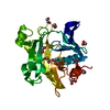
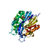
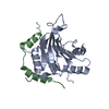
| Entry | Database: PDB / ID: 3ouj | ||||||
|---|---|---|---|---|---|---|---|
| Title | PHD2 with 2-Oxoglutarate | ||||||
 Components Components | Egl nine homolog 1 | ||||||
 Keywords Keywords |  OXIDOREDUCTASE / PHD2 / 2OG OXIDOREDUCTASE / PHD2 / 2OG | ||||||
| Function / homology |  Function and homology informationhypoxia-inducible factor-proline dioxygenase activity / hypoxia-inducible factor-proline dioxygenase / peptidyl-proline 4-dioxygenase activity / peptidyl-proline dioxygenase activity / peptidyl-proline hydroxylation to 4-hydroxy-L-proline / negative regulation of cyclic-nucleotide phosphodiesterase activity / regulation protein catabolic process at postsynapse / intracellular oxygen homeostasis / labyrinthine layer development / 2-oxoglutarate-dependent dioxygenase activity ...hypoxia-inducible factor-proline dioxygenase activity / hypoxia-inducible factor-proline dioxygenase / peptidyl-proline 4-dioxygenase activity / peptidyl-proline dioxygenase activity / peptidyl-proline hydroxylation to 4-hydroxy-L-proline / negative regulation of cyclic-nucleotide phosphodiesterase activity / regulation protein catabolic process at postsynapse / intracellular oxygen homeostasis / labyrinthine layer development / 2-oxoglutarate-dependent dioxygenase activity / cardiac muscle tissue morphogenesis / heart trabecula formation / regulation of modification of postsynaptic structure / L-ascorbic acid binding / response to nitric oxide / ventricular septum morphogenesis / regulation of angiogenesis / ferrous iron binding / Oxygen-dependent proline hydroxylation of Hypoxia-inducible Factor Alpha / negative regulation of DNA-binding transcription factor activity / cellular response to hypoxia / intracellular iron ion homeostasis / postsynaptic density / response to hypoxia / intracellular membrane-bounded organelle / glutamatergic synapse / enzyme binding / positive regulation of transcription by RNA polymerase II / nucleus / cytosol / cytoplasm Function and homology informationhypoxia-inducible factor-proline dioxygenase activity / hypoxia-inducible factor-proline dioxygenase / peptidyl-proline 4-dioxygenase activity / peptidyl-proline dioxygenase activity / peptidyl-proline hydroxylation to 4-hydroxy-L-proline / negative regulation of cyclic-nucleotide phosphodiesterase activity / regulation protein catabolic process at postsynapse / intracellular oxygen homeostasis / labyrinthine layer development / 2-oxoglutarate-dependent dioxygenase activity ...hypoxia-inducible factor-proline dioxygenase activity / hypoxia-inducible factor-proline dioxygenase / peptidyl-proline 4-dioxygenase activity / peptidyl-proline dioxygenase activity / peptidyl-proline hydroxylation to 4-hydroxy-L-proline / negative regulation of cyclic-nucleotide phosphodiesterase activity / regulation protein catabolic process at postsynapse / intracellular oxygen homeostasis / labyrinthine layer development / 2-oxoglutarate-dependent dioxygenase activity / cardiac muscle tissue morphogenesis / heart trabecula formation / regulation of modification of postsynaptic structure / L-ascorbic acid binding / response to nitric oxide / ventricular septum morphogenesis / regulation of angiogenesis / ferrous iron binding / Oxygen-dependent proline hydroxylation of Hypoxia-inducible Factor Alpha / negative regulation of DNA-binding transcription factor activity / cellular response to hypoxia / intracellular iron ion homeostasis / postsynaptic density / response to hypoxia / intracellular membrane-bounded organelle / glutamatergic synapse / enzyme binding / positive regulation of transcription by RNA polymerase II / nucleus / cytosol / cytoplasmSimilarity search - Function | ||||||
| Biological species |  Homo sapiens (human) Homo sapiens (human) | ||||||
| Method | X-RAY DIFFRACTION / SYNCHROTRON / MOLECULAR REPLACEMENT / Resolution: 2.3 Å | ||||||
 Authors Authors | Staker, B.L. / Arakaki, T.L. | ||||||
 Citation Citation | Journal: ACS Med Chem Lett / Year: 2010 Title: Benzimidazole-2-pyrazole HIF Prolyl 4-Hydroxylase Inhibitors as Oral Erythropoietin Secretagogues. Authors: Rosen, M.D. / Venkatesan, H. / Peltier, H.M. / Bembenek, S.D. / Kanelakis, K.C. / Zhao, L.X. / Leonard, B.E. / Hocutt, F.M. / Wu, X. / Palomino, H.L. / Brondstetter, T.I. / Haugh, P.V. / ...Authors: Rosen, M.D. / Venkatesan, H. / Peltier, H.M. / Bembenek, S.D. / Kanelakis, K.C. / Zhao, L.X. / Leonard, B.E. / Hocutt, F.M. / Wu, X. / Palomino, H.L. / Brondstetter, T.I. / Haugh, P.V. / Cagnon, L. / Yan, W. / Liotta, L.A. / Young, A. / Mirzadegan, T. / Shankley, N.P. / Barrett, T.D. / Rabinowitz, M.H. | ||||||
| History |
|
- Structure visualization
| Structure viewer | Molecule: MolmilJmol/JSmol |
|---|
- Downloads & links
-Download
| PDBx/mmCIF format | 3ouj.cif.gz | 60.1 KB | Display | PDBx/mmCIF format |
|---|---|---|---|---|
| PDB format | pdb3ouj.ent.gz | 42.8 KB | Display | PDB format |
| PDBx/mmJSON format | 3ouj.json.gz | Tree view | PDBx/mmJSON format | |
| Others |  Other downloads Other downloads |
-Validation report
| Arichive directory | https://data.pdbj.org/pub/pdb/validation_reports/ou/3oujftp://data.pdbj.org/pub/pdb/validation_reports/ou/3ouj | HTTPS FTP |
|---|
-Related structure data


-Links
 PDBj
PDBj


- Assembly
| Deposited unit | 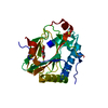
| ||||||||
|---|---|---|---|---|---|---|---|---|---|
| 1 |
| ||||||||
| Unit cell |
|
-Components
| #1: Protein | Mass: 26579.275 Da / Num. of mol.: 1 Source method: isolated from a genetically manipulated source Source: (gene. exp.) Homo sapiens (human) / Gene: EGLN1, C1orf12, PNAS-118, PNAS-137 / Plasmid: PBAD-SMT / Production host:  ESCHERICHIA COLI (E. coli) / Strain (production host): BL21(DE3) ESCHERICHIA COLI (E. coli) / Strain (production host): BL21(DE3)References: UniProt: Q9GZT9, Oxidoreductases; Acting on paired donors, with incorporation or reduction of molecular oxygen; With 2-oxoglutarate as one donor, and incorporation of one atom of oxygen into each donor |
|---|---|
| #2: Chemical | ChemComp-SO4 / Sulfate  Mass: 96.063 Da / Num. of mol.: 1 / Source method: obtained synthetically / Formula: SO4 Mass: 96.063 Da / Num. of mol.: 1 / Source method: obtained synthetically / Formula: SO4 |
| #3: Chemical | ChemComp-FE2 /   Mass: 55.845 Da / Num. of mol.: 1 / Source method: obtained synthetically / Formula: Fe Mass: 55.845 Da / Num. of mol.: 1 / Source method: obtained synthetically / Formula: Fe |
| #4: Chemical | ChemComp-AKG / Α-Ketoglutaric acid  Mass: 146.098 Da / Num. of mol.: 1 / Source method: obtained synthetically / Formula: C5H6O5 Mass: 146.098 Da / Num. of mol.: 1 / Source method: obtained synthetically / Formula: C5H6O5 |
| #5: Water | ChemComp-HOH / Water Mass: 18.015 Da / Num. of mol.: 53 / Source method: isolated from a natural source / Formula: H2O Mass: 18.015 Da / Num. of mol.: 53 / Source method: isolated from a natural source / Formula: H2O |
-Experimental details
-Experiment
| Experiment | Method: X-RAY DIFFRACTION / Number of used crystals: 1 |
|---|
- Sample preparation
| Crystal | Density Matthews: 2.29 Å3/Da / Density % sol: 46.19 % |
|---|---|
| Crystal grow | Temperature: 293 K / Method: vapor diffusion, sitting drop / pH: 6.4 Details: 0.2 M AMMONIUM SULFATE, 28% PEG 8000, pH 6.4, VAPOR DIFFUSION, SITTING DROP, temperature 293K |
-Data collection
| Diffraction | Mean temperature: 100 K |
|---|---|
| Diffraction source | Source: SYNCHROTRON / Site: APS  / Beamline: 23-ID-D / Wavelength: 1 / Beamline: 23-ID-D / Wavelength: 1 |
| Detector | Date: Nov 29, 2006 |
| Radiation | Protocol: SINGLE WAVELENGTH / Monochromatic (M) / Laue (L): M / Scattering type: x-ray |
| Radiation wavelength | Wavelength: 1 Å / Relative weight: 1 |
| Reflection | Resolution: 2.3→50 Å / Num. obs: 10691 / % possible obs: 99.1 % / Redundancy: 3.7 % / Rmerge(I) obs: 0.113 / Net I/σ(I): 6.1 |
| Reflection shell | Resolution: 2.3→2.38 Å / Redundancy: 2.6 % / Rmerge(I) obs: 0.33 / Mean I/σ(I) obs: 2.9 / % possible all: 95.1 |
- Processing
| Software | Name: REFMAC / Version: 5.5.0109 / Classification: refinement | ||||||||||||||||||||||||||||||||||||||||||||||||||||||||||||||||||||||||||||||||||||||||||||||||||||||||||||||||||||||||||||||||||||||||||||||||||||||||||||||||||||||||||
|---|---|---|---|---|---|---|---|---|---|---|---|---|---|---|---|---|---|---|---|---|---|---|---|---|---|---|---|---|---|---|---|---|---|---|---|---|---|---|---|---|---|---|---|---|---|---|---|---|---|---|---|---|---|---|---|---|---|---|---|---|---|---|---|---|---|---|---|---|---|---|---|---|---|---|---|---|---|---|---|---|---|---|---|---|---|---|---|---|---|---|---|---|---|---|---|---|---|---|---|---|---|---|---|---|---|---|---|---|---|---|---|---|---|---|---|---|---|---|---|---|---|---|---|---|---|---|---|---|---|---|---|---|---|---|---|---|---|---|---|---|---|---|---|---|---|---|---|---|---|---|---|---|---|---|---|---|---|---|---|---|---|---|---|---|---|---|---|---|---|---|---|
| Refinement | Method to determine structure: MOLECULAR REPLACEMENT / Resolution: 2.3→32.33 Å / Cor.coef. Fo:Fc: 0.929 / Cor.coef. Fo:Fc free: 0.888 / SU B: 7.357 / SU ML: 0.188 / Cross valid method: THROUGHOUT / σ(F): 0 / ESU R Free: 0.255 / Stereochemistry target values: MAXIMUM LIKELIHOOD
| ||||||||||||||||||||||||||||||||||||||||||||||||||||||||||||||||||||||||||||||||||||||||||||||||||||||||||||||||||||||||||||||||||||||||||||||||||||||||||||||||||||||||||
| Solvent computation | Ion probe radii: 0.8 Å / Shrinkage radii: 0.8 Å / VDW probe radii: 1.4 Å / Solvent model: BABINET MODEL WITH MASK | ||||||||||||||||||||||||||||||||||||||||||||||||||||||||||||||||||||||||||||||||||||||||||||||||||||||||||||||||||||||||||||||||||||||||||||||||||||||||||||||||||||||||||
| Displacement parameters | Biso mean: 33.83 Å2
| ||||||||||||||||||||||||||||||||||||||||||||||||||||||||||||||||||||||||||||||||||||||||||||||||||||||||||||||||||||||||||||||||||||||||||||||||||||||||||||||||||||||||||
| Refinement step | Cycle: LAST / Resolution: 2.3→32.33 Å
| ||||||||||||||||||||||||||||||||||||||||||||||||||||||||||||||||||||||||||||||||||||||||||||||||||||||||||||||||||||||||||||||||||||||||||||||||||||||||||||||||||||||||||
| Refine LS restraints |
| ||||||||||||||||||||||||||||||||||||||||||||||||||||||||||||||||||||||||||||||||||||||||||||||||||||||||||||||||||||||||||||||||||||||||||||||||||||||||||||||||||||||||||
| LS refinement shell | Resolution: 2.3→2.36 Å / Total num. of bins used: 20
|
