- PDB-3orj: Crystal structure of a sugar-binding protein (BACOVA_04391) from ... -
+
Open data
ID or keywords:
Loading...
-
Basic information
Entry
Database: PDB / ID: 3orj
Title
Crystal structure of a sugar-binding protein (BACOVA_04391) from Bacteroides ovatus at 2.16 A resolution
Components
sugar-binding protein
Keywords
SUGAR BINDING PROTEIN / STRUCTURAL GENOMICS / JOINT CENTER FOR STRUCTURAL GENOMICS / JCSG / PROTEIN STRUCTURE INITIATIVE / PSI-BIOLOGY / SUGAR-BINDING PROTEIN
Function / homology
Surface glycan-binding protein B, xyloglucan binding domain / Surface glycan-binding protein B xyloglucan binding domain / polysaccharide binding / Immunoglobulins / Immunoglobulin-like fold / Immunoglobulin-like / Sandwich / Mainly Beta / SGBP_B_XBD domain-containing protein
Function and homology information
Biological species
Bacteroides ovatus (bacteria)
Method
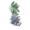
X-RAY DIFFRACTION / SYNCHROTRON / MAD / Resolution: 2.16 Å
ANALYTICAL SIZE EXCLUSION CHROMATOGRAPHY WITH STATIC LIGHT SCATTERING SUPPORTS THE ASSIGNMENT OF A MONOMER AS A SIGNIFICANT OLIGOMERIZATION STATE IN SOLUTION.
-
Components
#1: Protein
sugar-bindingprotein
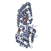
Mass: 48137.301 Da / Num. of mol.: 1 Source method: isolated from a genetically manipulated source Source: (gene. exp.) Bacteroides ovatus (bacteria) / Strain: ATCC 8483 / Gene: BACOVA_04391 / Plasmid: SpeedET / Production host: Escherichia Coli (E. coli) / Strain (production host): HK100 / References: UniProt: A7M2Q4
Mass: 18.015 Da / Num. of mol.: 275 / Source method: isolated from a natural source / Formula: H2O
Sequence details
THE CONSTRUCT (RESIDUES 30-467) WAS EXPRESSED WITH A PURIFICATION TAG MGSDKIHHHHHHENLYFQG. THE TAG ...THE CONSTRUCT (RESIDUES 30-467) WAS EXPRESSED WITH A PURIFICATION TAG MGSDKIHHHHHHENLYFQG. THE TAG WAS REMOVED WITH TEV PROTEASE LEAVING ONLY A GLYCINE (0) FOLLOWED BY THE TARGET SEQUENCE.
-
Experimental details
-
Experiment
Experiment
Method: X-RAY DIFFRACTION / Number of used crystals: 1
-
Sample preparation
Crystal
Density Matthews: 2.64 Å3/Da / Density % sol: 53.4 %
Crystal grow
Temperature: 277 K / Method: vapor diffusion, sitting drop / pH: 9 Details: 30.0% PEG-6000, 0.1M Bicine pH 9.0, NANODROP, VAPOR DIFFUSION, SITTING DROP, temperature 277K
Type: MARMOSAIC 325 mm CCD / Detector: CCD / Date: Jun 9, 2010 / Details: Flat mirror (vertical focusing)
Radiation
Monochromator: Single crystal Si(111) bent monochromator (horizontal focusing) Protocol: MAD / Monochromatic (M) / Laue (L): M / Scattering type: x-ray
Radiation wavelength
ID
Wavelength (Å)
Relative weight
1
0.93925
1
2
0.97939
1
3
0.97891
1
Reflection
Resolution: 2.16→45.372 Å / Num. obs: 26772 / % possible obs: 95.2 % / Observed criterion σ(I): -3 / Redundancy: 3.55 % / Biso Wilson estimate: 42.666 Å2 / Rmerge(I) obs: 0.047 / Net I/σ(I): 16.67
Reflection shell
Resolution (Å)
Rmerge(I) obs
Mean I/σ(I) obs
Num. measured obs
Num. unique obs
Diffraction-ID
% possible all
2.16-2.24
0.614
2
6299
2004
1
70.4
2.24-2.33
0.408
3.2
8406
2483
1
90.5
2.33-2.43
0.305
4
9525
2592
1
99
2.43-2.56
0.238
5.1
10269
2784
1
99
2.56-2.72
0.165
7.1
10024
2748
1
99.3
2.72-2.93
0.105
10.8
10167
2782
1
99
2.93-3.22
0.061
17.3
10015
2742
1
99.4
3.22-3.69
0.035
27.6
10108
2821
1
98.7
3.69-4.64
0.025
38.2
10055
2822
1
99
4.64-45.37
0.024
42.6
10136
2977
1
97.9
-
Phasing
Phasing
Method: MAD
-
Processing
Software
Name
Version
Classification
NB
SHELX
phasing
BUSTER-TNT
BUSTER2.8.0
refinement
XSCALE
dataprocessing
PDB_EXTRACT
3.1
dataextraction
XDS
datareduction
XSCALE
datascaling
SHELXD
phasing
autoSHARP
phasing
BUSTER
2.8.0
refinement
Refinement
Method to determine structure: MAD / Resolution: 2.16→45.372 Å / Cor.coef. Fo:Fc: 0.9377 / Cor.coef. Fo:Fc free: 0.9235 / Occupancy max: 1 / Occupancy min: 0.5 / Cross valid method: THROUGHOUT / σ(F): 0 Details: 1. A MET-INHIBITION PROTOCOL WAS USED FOR SELENOMETHIONINE INCORPORATION DURING PROTEIN EXPRESSION. THE OCCUPANCY OF THE SE ATOMS IN THE MSE RESIDUES WAS REDUCED TO 0.75 FOR THE REDUCED ...Details: 1. A MET-INHIBITION PROTOCOL WAS USED FOR SELENOMETHIONINE INCORPORATION DURING PROTEIN EXPRESSION. THE OCCUPANCY OF THE SE ATOMS IN THE MSE RESIDUES WAS REDUCED TO 0.75 FOR THE REDUCED SCATTERING POWER DUE TO PARTIAL S-MET INCORPORATION. 2. CHLORIDE (CL) MODELED IS PRESENT PROTEIN BUFFER. ETHYLENE GLYCOL (EDO) MODELED ARE PRESENT CRYO SOLUTION. 3. ZINC ION IS TENTATIVELY ASSIGNED BASED ON ANOMALOUS DIFFERENCE MAP AND GEOMETRY.
In the structure databanks used in Yorodumi, some data are registered as the other names, "COVID-19 virus" and "2019-nCoV". Here are the details of the virus and the list of structure data.
Jan 31, 2019. EMDB accession codes are about to change! (news from PDBe EMDB page)
EMDB accession codes are about to change! (news from PDBe EMDB page)
The allocation of 4 digits for EMDB accession codes will soon come to an end. Whilst these codes will remain in use, new EMDB accession codes will include an additional digit and will expand incrementally as the available range of codes is exhausted. The current 4-digit format prefixed with “EMD-” (i.e. EMD-XXXX) will advance to a 5-digit format (i.e. EMD-XXXXX), and so on. It is currently estimated that the 4-digit codes will be depleted around Spring 2019, at which point the 5-digit format will come into force.
The EM Navigator/Yorodumi systems omit the EMD- prefix.
Related info.:Q: What is EMD? / ID/Accession-code notation in Yorodumi/EM Navigator
Yorodumi is a browser for structure data from EMDB, PDB, SASBDB, etc.
This page is also the successor to EM Navigator detail page, and also detail information page/front-end page for Omokage search.
The word "yorodu" (or yorozu) is an old Japanese word meaning "ten thousand". "mi" (miru) is to see.
Related info.:EMDB / PDB / SASBDB / Comparison of 3 databanks / Yorodumi Search / Aug 31, 2016. New EM Navigator & Yorodumi / Yorodumi Papers / Jmol/JSmol / Function and homology information / Changes in new EM Navigator and Yorodumi
 Movie
Movie Controller

 Yorodumi
 Open data
 Basic information
 Components
 Keywords
 STRUCTURAL GENOMICS / JOINT CENTER FOR STRUCTURAL GENOMICS / JCSG /
  Function and homology information
 Bacteroides ovatus (bacteria)
 Authors
 Citation
 Structure visualization
 Downloads & links
 Other downloads
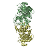


 PDBj
PDBj Assembly

 Mass: 65.409 Da / Num. of mol.: 1 / Source method: obtained synthetically / Formula: Zn
 Mass: 35.453 Da / Num. of mol.: 1 / Source method: obtained synthetically / Formula: Cl
 Mass: 62.068 Da / Num. of mol.: 4 / Source method: obtained synthetically / Formula: C2H6O2
 Mass: 18.015 Da / Num. of mol.: 275 / Source method: isolated from a natural source / Formula: H2O
 Sample preparation
 / Beamline: BL11-1 / Wavelength: 0.93925,0.97939,0.97891
 Processing