 Movie
Movie Controller

[English] 日本語
 Yorodumi
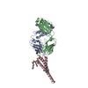
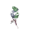
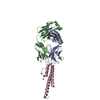
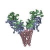
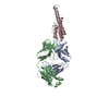
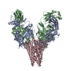
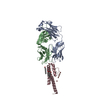
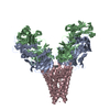
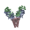
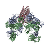
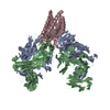
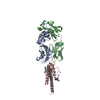
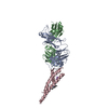
- PDB-3or7: On the structural basis of modal gating behavior in K+channels - E71I -
+ Open data

- Basic information
| Entry | Database: PDB / ID: 3or7 | ||||||
|---|---|---|---|---|---|---|---|
| Title | On the structural basis of modal gating behavior in K+channels - E71I | ||||||
 Components Components |
| ||||||
 Keywords Keywords |  IMMUNE SYSTEM/TRANSPORT PROTEIN / inactivation / alpha-helical / Potassium channel / IMMUNE SYSTEM-TRANSPORT PROTEIN complex IMMUNE SYSTEM/TRANSPORT PROTEIN / inactivation / alpha-helical / Potassium channel / IMMUNE SYSTEM-TRANSPORT PROTEIN complex | ||||||
| Function / homology |  Function and homology information Function and homology informationmonoatomic ion transmembrane transport / identical protein binding / plasma membraneSimilarity search - Function | ||||||
| Biological species |  Streptomyces lividans (bacteria) Streptomyces lividans (bacteria) Mus musculus (house mouse) Mus musculus (house mouse) | ||||||
| Method | X-RAY DIFFRACTION / SYNCHROTRON / MOLECULAR REPLACEMENT / Resolution: 2.3 Å | ||||||
 Authors Authors | Chakrapani, S. / Cordero-Morales, J.F. / Jogini, V. / Pan, A.C. / Cortes, D.M. / Roux, B. / Perozo, E. | ||||||
 Citation Citation | Journal: Nat.Struct.Mol.Biol. / Year: 2011 Title: On the structural basis of modal gating behavior in K(+) channels. Authors: Chakrapani, S. / Cordero-Morales, J.F. / Jogini, V. / Pan, A.C. / Cortes, D.M. / Roux, B. / Perozo, E. | ||||||
| History |
|
- Structure visualization
| Structure viewer | Molecule: MolmilJmol/JSmol |
|---|
- Downloads & links
-Download
| PDBx/mmCIF format | 3or7.cif.gz | 109 KB | Display | PDBx/mmCIF format |
|---|---|---|---|---|
| PDB format | pdb3or7.ent.gz | 88.1 KB | Display | PDB format |
| PDBx/mmJSON format | 3or7.json.gz | Tree view | PDBx/mmJSON format | |
| Others |  Other downloads Other downloads |
-Validation report
| Arichive directory | https://data.pdbj.org/pub/pdb/validation_reports/or/3or7ftp://data.pdbj.org/pub/pdb/validation_reports/or/3or7 | HTTPS FTP |
|---|
-Related structure data


-Links
 PDBj
PDBj


- Assembly
| Deposited unit | 
| ||||||||||||||||||||||||
|---|---|---|---|---|---|---|---|---|---|---|---|---|---|---|---|---|---|---|---|---|---|---|---|---|---|
| 1 | 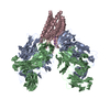
| ||||||||||||||||||||||||
| Unit cell |
| ||||||||||||||||||||||||
| Components on special symmetry positions |
|
-Components
| #1: Antibody | Mass: 23411.242 Da / Num. of mol.: 1 / Source method: isolated from a natural source / Source: (natural) Mus musculus (house mouse) | ||
|---|---|---|---|
| #2: Antibody | Mass: 23435.738 Da / Num. of mol.: 1 / Source method: isolated from a natural source / Source: (natural) Mus musculus (house mouse) | ||
| #3: Protein | Mass: 10962.780 Da / Num. of mol.: 1 / Fragment: UNP RESIDUES 22-124 / Mutation: E71I Source method: isolated from a genetically manipulated source Source: (gene. exp.) Streptomyces lividans (bacteria) / Production host: Escherichia coli (E. coli) / References: UniProt: P0A334 | ||
| #4: Chemical | ChemComp-K /   Mass: 39.098 Da / Num. of mol.: 5 / Source method: obtained synthetically / Formula: K Mass: 39.098 Da / Num. of mol.: 5 / Source method: obtained synthetically / Formula: K#5: Water | ChemComp-HOH / | Water Mass: 18.015 Da / Num. of mol.: 7 / Source method: isolated from a natural source / Formula: H2O Mass: 18.015 Da / Num. of mol.: 7 / Source method: isolated from a natural source / Formula: H2O |
-Experimental details
-Experiment
| Experiment | Method: X-RAY DIFFRACTION / Number of used crystals: 1 |
|---|
- Sample preparation
| Crystal | Density Matthews: 4.02 Å3/Da / Density % sol: 69.4 % |
|---|---|
| Crystal grow | Temperature: 298 K / Method: vapor diffusion, sitting drop / pH: 6 Details: 20-25% PEG400, 50mM magnesium acetate, 50mM sodium acetate pH 5.0-6.0, VAPOR DIFFUSION, SITTING DROP, temperature 298K |
-Data collection
| Diffraction source | Source: SYNCHROTRON / Site: APS  / Beamline: 19-ID / Wavelength: 1 Å / Beamline: 19-ID / Wavelength: 1 Å |
|---|---|
| Detector | Type: ADSC QUANTUM 315 / Detector: CCD / Date: Oct 13, 2006 |
| Radiation | Protocol: SINGLE WAVELENGTH / Monochromatic (M) / Laue (L): M / Scattering type: x-ray |
| Radiation wavelength | Wavelength: 1 Å / Relative weight: 1 |
| Reflection | Resolution: 2.3→40 Å / Num. all: 40893 / Num. obs: 39318 / % possible obs: 96 % / Observed criterion σ(F): 2 / Observed criterion σ(I): 2 |
- Processing
| Software |
| |||||||||||||||
|---|---|---|---|---|---|---|---|---|---|---|---|---|---|---|---|---|
| Refinement | Method to determine structure: MOLECULAR REPLACEMENT / Resolution: 2.3→40 Å / σ(F): 2
| |||||||||||||||
| Refinement step | Cycle: LAST / Resolution: 2.3→40 Å
| |||||||||||||||
| Refine LS restraints |
|
