 Movie
Movie Controller

[English] 日本語
 Yorodumi
- PDB-3op7: Crystal structure of a PLP-dependent aminotransferase (ZP_0362512... -
+ Open data

- Basic information
| Entry | Database: PDB / ID: 3op7 | ||||||
|---|---|---|---|---|---|---|---|
| Title | Crystal structure of a PLP-dependent aminotransferase (ZP_03625122.1) from Streptococcus suis 89-1591 at 1.70 A resolution | ||||||
 Components Components | Aminotransferase class I and II | ||||||
 Keywords Keywords |  TRANSFERASE / PLP-DEPENDENT TRANSFERASE / STRUCTURAL GENOMICS / JOINT CENTER FOR STRUCTURAL GENOMICS / JCSG / PROTEIN STRUCTURE INITIATIVE / PSI-BIOLOGY TRANSFERASE / PLP-DEPENDENT TRANSFERASE / STRUCTURAL GENOMICS / JOINT CENTER FOR STRUCTURAL GENOMICS / JCSG / PROTEIN STRUCTURE INITIATIVE / PSI-BIOLOGY | ||||||
| Function / homology | Aspartate Aminotransferase, domain 1 / Aspartate Aminotransferase, domain 1 / Aspartate Aminotransferase; domain 2 / Type I PLP-dependent aspartate aminotransferase-like (Major domain) / Alpha-Beta Complex / 3-Layer(aba) Sandwich / Alpha Beta / Unknown ligand / :  Function and homology information Function and homology information | ||||||
| Biological species |  Streptococcus suis 89/1591 (bacteria) Streptococcus suis 89/1591 (bacteria) | ||||||
| Method | X-RAY DIFFRACTION / SYNCHROTRON / SAD / Resolution: 1.7 Å | ||||||
 Authors Authors | Joint Center for Structural Genomics (JCSG) | ||||||
 Citation Citation | Journal: To be published Title: Crystal structure of a PLP-dependent aminotransferase (ZP_03625122.1) from Streptococcus suis 89-1591 at 1.70 A resolution Authors: Joint Center for Structural Genomics (JCSG) | ||||||
| History |
|
- Structure visualization
| Structure viewer | Molecule: MolmilJmol/JSmol |
|---|
- Downloads & links
-Download
| PDBx/mmCIF format | 3op7.cif.gz | 169.5 KB | Display | PDBx/mmCIF format |
|---|---|---|---|---|
| PDB format | pdb3op7.ent.gz | 137.5 KB | Display | PDB format |
| PDBx/mmJSON format | 3op7.json.gz | Tree view | PDBx/mmJSON format | |
| Others |  Other downloads Other downloads |
-Validation report
| Arichive directory | https://data.pdbj.org/pub/pdb/validation_reports/op/3op7ftp://data.pdbj.org/pub/pdb/validation_reports/op/3op7 | HTTPS FTP |
|---|
-Related structure data
| Similar structure data | |
|---|---|
| Other databases |


-Links
 PDBj
PDBj

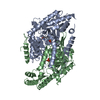
- Assembly
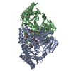
| Deposited unit | 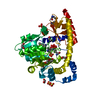
| ||||||||
|---|---|---|---|---|---|---|---|---|---|
| 1 | 
| ||||||||
| Unit cell |
| ||||||||
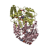
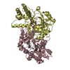
| Details | CRYSTAL PACKING ANALYSIS SUGGESTS THE ASSIGNMENT OF A DIMER AS THE SIGNIFICANT OLIGOMERIZATION STATE. |
-Components
| #1: Protein | Mass: 43039.711 Da / Num. of mol.: 1 Source method: isolated from a genetically manipulated source Source: (gene. exp.) Streptococcus suis 89/1591 (bacteria) / Gene: SsuiDRAFT_2941 / Plasmid: SpeedET / Production host: Escherichia Coli (E. coli) / Strain (production host): HK100 / References: UniProt: B9WVA1 | ||||||
|---|---|---|---|---|---|---|---|
| #2: Chemical | ChemComp-UNL / Num. of mol.: 1 / Source method: obtained synthetically | ||||||
| #3: Chemical | Sulfate  Mass: 96.063 Da / Num. of mol.: 3 / Source method: obtained synthetically / Formula: SO4 Mass: 96.063 Da / Num. of mol.: 3 / Source method: obtained synthetically / Formula: SO4#4: Chemical | ChemComp-EDO / Ethylene glycol  Mass: 62.068 Da / Num. of mol.: 6 / Source method: obtained synthetically / Formula: C2H6O2 Mass: 62.068 Da / Num. of mol.: 6 / Source method: obtained synthetically / Formula: C2H6O2#5: Water | ChemComp-HOH / | Water Mass: 18.015 Da / Num. of mol.: 277 / Source method: isolated from a natural source / Formula: H2O Mass: 18.015 Da / Num. of mol.: 277 / Source method: isolated from a natural source / Formula: H2OSequence details | THIS CONSTRUCT WAS EXPRESSED WITH A PURIFICATION TAG MGSDKIHHHHHHENLYFQG. THE TAG WAS REMOVED WITH ...THIS CONSTRUCT WAS EXPRESSED WITH A PURIFICATI | |
-Experimental details
-Experiment
| Experiment | Method: X-RAY DIFFRACTION / Number of used crystals: 1 |
|---|
- Sample preparation
| Crystal | Density Matthews: 2.46 Å3/Da / Density % sol: 50.1 % |
|---|---|
| Crystal grow | Temperature: 277 K / Method: vapor diffusion, sitting drop Details: 0.20M lithium sulfate, 2.00M ammonium sulfate, Additive: 0.001M pyridoxal 5'-phosphate (PLP), NANODROP, VAPOR DIFFUSION, SITTING DROP, temperature 277K |
-Data collection
| Diffraction | Mean temperature: 100 K | ||||||||||||||||||||||||||||||||||||||||||||||||||||||||||||||||||||||||||||||||||||||||||||||||||||||||||||||||||||||||||||||||||||||||||||||||||||||||||||||||||||||||
|---|---|---|---|---|---|---|---|---|---|---|---|---|---|---|---|---|---|---|---|---|---|---|---|---|---|---|---|---|---|---|---|---|---|---|---|---|---|---|---|---|---|---|---|---|---|---|---|---|---|---|---|---|---|---|---|---|---|---|---|---|---|---|---|---|---|---|---|---|---|---|---|---|---|---|---|---|---|---|---|---|---|---|---|---|---|---|---|---|---|---|---|---|---|---|---|---|---|---|---|---|---|---|---|---|---|---|---|---|---|---|---|---|---|---|---|---|---|---|---|---|---|---|---|---|---|---|---|---|---|---|---|---|---|---|---|---|---|---|---|---|---|---|---|---|---|---|---|---|---|---|---|---|---|---|---|---|---|---|---|---|---|---|---|---|---|---|---|---|---|
| Diffraction source | Source: SYNCHROTRON / Site: SSRL  / Beamline: BL9-2 / Wavelength: 0.97905 / Beamline: BL9-2 / Wavelength: 0.97905 | ||||||||||||||||||||||||||||||||||||||||||||||||||||||||||||||||||||||||||||||||||||||||||||||||||||||||||||||||||||||||||||||||||||||||||||||||||||||||||||||||||||||||
| Detector | Type: MARMOSAIC 325 mm CCD / Detector: CCD / Date: Jul 23, 2010 / Details: Flat collimating mirror, toroid focusing mirror | ||||||||||||||||||||||||||||||||||||||||||||||||||||||||||||||||||||||||||||||||||||||||||||||||||||||||||||||||||||||||||||||||||||||||||||||||||||||||||||||||||||||||
| Radiation | Monochromator: Double crystal monochromator / Protocol: SINGLE WAVELENGTH / Monochromatic (M) / Laue (L): M / Scattering type: x-ray | ||||||||||||||||||||||||||||||||||||||||||||||||||||||||||||||||||||||||||||||||||||||||||||||||||||||||||||||||||||||||||||||||||||||||||||||||||||||||||||||||||||||||
| Radiation wavelength | Wavelength: 0.97905 Å / Relative weight: 1 | ||||||||||||||||||||||||||||||||||||||||||||||||||||||||||||||||||||||||||||||||||||||||||||||||||||||||||||||||||||||||||||||||||||||||||||||||||||||||||||||||||||||||
| Reflection | Resolution: 1.7→29.748 Å / Num. all: 47906 / Num. obs: 47906 / % possible obs: 100 % / Redundancy: 7.3 % / Biso Wilson estimate: 24.986 Å2 / Rsym value: 0.087 / Net I/σ(I): 11.2 | ||||||||||||||||||||||||||||||||||||||||||||||||||||||||||||||||||||||||||||||||||||||||||||||||||||||||||||||||||||||||||||||||||||||||||||||||||||||||||||||||||||||||
| Reflection shell | Diffraction-ID: 1
|
-Phasing
| Phasing | Method: SAD |
|---|
- Processing
| Software |
| |||||||||||||||||||||||||||||||||||||||||||||||||||||||||||||||||||||||||||||||||||||
|---|---|---|---|---|---|---|---|---|---|---|---|---|---|---|---|---|---|---|---|---|---|---|---|---|---|---|---|---|---|---|---|---|---|---|---|---|---|---|---|---|---|---|---|---|---|---|---|---|---|---|---|---|---|---|---|---|---|---|---|---|---|---|---|---|---|---|---|---|---|---|---|---|---|---|---|---|---|---|---|---|---|---|---|---|---|---|
| Refinement | Method to determine structure: SAD / Resolution: 1.7→29.748 Å / Cor.coef. Fo:Fc: 0.971 / Cor.coef. Fo:Fc free: 0.959 / Occupancy max: 1 / Occupancy min: 0.3 / SU B: 4.15 / SU ML: 0.067 / Cross valid method: THROUGHOUT / σ(F): 0 / ESU R: 0.092 / ESU R Free: 0.093 Stereochemistry target values: MAXIMUM LIKELIHOOD WITH PHASES Details: 1. HYDROGENS HAVE BEEN ADDED IN THE RIDING POSITIONS. 2. ATOM RECORD CONTAINS SUM OF TLS AND RESIDUAL B FACTORS. 3. ANISOU RECORD CONTAINS SUM OF TLS AND RESIDUAL U FACTORS. 4. WATERS WERE ...Details: 1. HYDROGENS HAVE BEEN ADDED IN THE RIDING POSITIONS. 2. ATOM RECORD CONTAINS SUM OF TLS AND RESIDUAL B FACTORS. 3. ANISOU RECORD CONTAINS SUM OF TLS AND RESIDUAL U FACTORS. 4. WATERS WERE EXCLUDED FROM AUTOMATIC TLS ASSIGNMENT. 5. A MET-INHIBITION PROTOCOL WAS USED FOR SELENOMETHIONINE INCORPORATION DURING PROTEIN EXPRESSION. THE OCCUPANCY OF THE SE ATOMS IN THE MSE RESIDUES WAS REDUCED TO 0.75 FOR THE REDUCED SCATTERING POWER DUE TO PARTIAL S-MET INCORPORATION. 6. SULFATE (SO4) AND 1,2-ETHANEDIOL (EDO) MOLECULES FROM THE CRYSTALLIZATION/CRYOPROTECTION SOLUTION ARE MODELED. 7. RESIDUE LYSINE 221 IS COVALENTLY ATTACHED TO PYRIDOXAL-5'-PHOSPHATE VIA SCHIFF BASE LINKAGE AND IS MODELED AS LLP. 8. AN UNKNOWN LIGAND (UNL) HAS BEEN MODELED NEAR RESIDUE LLP 221. THE UNL RESEMBLES PYRIDOXAMINE (PXM), A POSSIBLE REACTION PRODUCT OF THE ENZYME.
| |||||||||||||||||||||||||||||||||||||||||||||||||||||||||||||||||||||||||||||||||||||
| Solvent computation | Ion probe radii: 0.8 Å / Shrinkage radii: 0.8 Å / VDW probe radii: 1.4 Å / Solvent model: BABINET MODEL WITH MASK | |||||||||||||||||||||||||||||||||||||||||||||||||||||||||||||||||||||||||||||||||||||
| Displacement parameters | Biso max: 86.44 Å2 / Biso mean: 35.7562 Å2 / Biso min: 17.78 Å2
| |||||||||||||||||||||||||||||||||||||||||||||||||||||||||||||||||||||||||||||||||||||
| Refinement step | Cycle: LAST / Resolution: 1.7→29.748 Å
| |||||||||||||||||||||||||||||||||||||||||||||||||||||||||||||||||||||||||||||||||||||
| Refine LS restraints |
| |||||||||||||||||||||||||||||||||||||||||||||||||||||||||||||||||||||||||||||||||||||
| LS refinement shell | Resolution: 1.7→1.744 Å / Total num. of bins used: 20
| |||||||||||||||||||||||||||||||||||||||||||||||||||||||||||||||||||||||||||||||||||||
| Refinement TLS params. | Method: refined / Origin x: 29.511 Å / Origin y: 28.9 Å / Origin z: 16.407 Å
|
