 Movie
Movie Controller

[English] 日本語
 Yorodumi
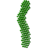
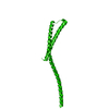
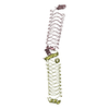
- PDB-3onx: Crystal structure of a domain of a protein involved in formation ... -
+ Open data

- Basic information
| Entry | Database: PDB / ID: 3onx | ||||||
|---|---|---|---|---|---|---|---|
| Title | Crystal structure of a domain of a protein involved in formation of actin cytoskeleton | ||||||
 Components Components | Bud site selection protein 6 | ||||||
 Keywords Keywords |  PROTEIN BINDING / coiled-coil PROTEIN BINDING / coiled-coil | ||||||
| Function / homology |  Function and homology information Function and homology informationcytoskeletal regulatory protein binding / positive regulation of formin-nucleated actin cable assembly / polarisome / bipolar cellular bud site selection / budding cell apical bud growth / secretory vesicle / vesicle targeting / pseudohyphal growth / prospore membrane / establishment or maintenance of actin cytoskeleton polarity ...cytoskeletal regulatory protein binding / positive regulation of formin-nucleated actin cable assembly / polarisome / bipolar cellular bud site selection / budding cell apical bud growth / secretory vesicle / vesicle targeting / pseudohyphal growth / prospore membrane / establishment or maintenance of actin cytoskeleton polarity / positive regulation of actin nucleation / astral microtubule organization / cell tip / incipient cellular bud site / cellular bud tip / cellular bud neck / mating projection tip / spindle pole body / establishment of cell polarity / actin filament bundle assembly / enzyme activator activity / regulation of actin cytoskeleton organization / regulation of protein localization / actin binding / cytoplasmSimilarity search - Function | ||||||
| Biological species |  Saccharomyces cerevisiae (brewer's yeast) Saccharomyces cerevisiae (brewer's yeast) | ||||||
| Method | X-RAY DIFFRACTION / SYNCHROTRON / SAD / Resolution: 2.904 Å | ||||||
 Authors Authors | Tu, D. / Eck, M.J. | ||||||
 Citation Citation | Journal: Proc.Natl.Acad.Sci.USA / Year: 2012 Title: Structure of the formin-interaction domain of the actin nucleation-promoting factor Bud6. Authors: Tu, D. / Graziano, B.R. / Park, E. / Zheng, W. / Li, Y. / Goode, B.L. / Eck, M.J. | ||||||
| History |
|
- Structure visualization
| Structure viewer | Molecule: MolmilJmol/JSmol |
|---|
- Downloads & links
-Download
| PDBx/mmCIF format | 3onx.cif.gz | 57.4 KB | Display | PDBx/mmCIF format |
|---|---|---|---|---|
| PDB format | pdb3onx.ent.gz | 46.9 KB | Display | PDB format |
| PDBx/mmJSON format | 3onx.json.gz | Tree view | PDBx/mmJSON format | |
| Others |  Other downloads Other downloads |
-Validation report
| Arichive directory | https://data.pdbj.org/pub/pdb/validation_reports/on/3onxftp://data.pdbj.org/pub/pdb/validation_reports/on/3onx | HTTPS FTP |
|---|
-Related structure data


-Links
 PDBj
PDBj- Assembly
| Deposited unit | 
| ||||||||
|---|---|---|---|---|---|---|---|---|---|
| 1 |
| ||||||||
| Unit cell |
|
-Components
| #1: Protein | Mass: 16496.176 Da / Num. of mol.: 2 / Fragment: coiled-coil domain (unp residues 549-688) Source method: isolated from a genetically manipulated source Source: (gene. exp.) Saccharomyces cerevisiae (brewer's yeast)Gene: AIP3, BUD6, L8543.5, YLR319C / Plasmid: pET30 / Production host:  Escherichia coli (E. coli) / Strain (production host): Rosetta2(DE3) / References: UniProt: P41697 Escherichia coli (E. coli) / Strain (production host): Rosetta2(DE3) / References: UniProt: P41697 |
|---|
-Experimental details
-Experiment
| Experiment | Method: X-RAY DIFFRACTION / Number of used crystals: 1 |
|---|
- Sample preparation
| Crystal | Density Matthews: 3.19 Å3/Da / Density % sol: 61.45 % |
|---|---|
| Crystal grow | Temperature: 293 K / Method: vapor diffusion, hanging drop / pH: 7 Details: 9% PEG 3350, 85 mM sodium malonate (pH 7.0), 25% glycerol, 10 mM DTT, VAPOR DIFFUSION, HANGING DROP, temperature 293K |
-Data collection
| Diffraction | Mean temperature: 100 K |
|---|---|
| Diffraction source | Source: SYNCHROTRON / Site: APS  / Beamline: 24-ID-C / Wavelength: 0.97934 Å / Beamline: 24-ID-C / Wavelength: 0.97934 Å |
| Detector | Type: ADSC QUANTUM 315 / Detector: CCD / Date: Jul 16, 2008 |
| Radiation | Monochromator: MD2 microdiffractometer / Protocol: SINGLE WAVELENGTH / Monochromatic (M) / Laue (L): M / Scattering type: x-ray |
| Radiation wavelength | Wavelength: 0.97934 Å / Relative weight: 1 |
| Reflection | Resolution: 2.9→50 Å / Num. obs: 17136 / % possible obs: 95.3 % / Observed criterion σ(F): 0 / Observed criterion σ(I): 0 / Redundancy: 1.9 % / Rmerge(I) obs: 0.044 / Net I/σ(I): 15.8 |
| Reflection shell | Resolution: 2.9→3 Å / Rmerge(I) obs: 0.282 / Mean I/σ(I) obs: 2 / Num. unique all: 1287 / % possible all: 71.7 |
- Processing
| Software |
| |||||||||||||||||||||||||||||||||||||||||||||||||||||||||||||||||||||||||||||||||||||||||||
|---|---|---|---|---|---|---|---|---|---|---|---|---|---|---|---|---|---|---|---|---|---|---|---|---|---|---|---|---|---|---|---|---|---|---|---|---|---|---|---|---|---|---|---|---|---|---|---|---|---|---|---|---|---|---|---|---|---|---|---|---|---|---|---|---|---|---|---|---|---|---|---|---|---|---|---|---|---|---|---|---|---|---|---|---|---|---|---|---|---|---|---|---|
| Refinement | Method to determine structure: SAD / Resolution: 2.904→35.028 Å / SU ML: 0.42 / Cross valid method: THROUGHOUT / σ(F): 0.7 / Phase error: 32.88 / Stereochemistry target values: MLHL
| |||||||||||||||||||||||||||||||||||||||||||||||||||||||||||||||||||||||||||||||||||||||||||
| Solvent computation | Shrinkage radii: 0.9 Å / VDW probe radii: 1.11 Å / Solvent model: FLAT BULK SOLVENT MODEL / Bsol: 65.01 Å2 / ksol: 0.323 e/Å3 | |||||||||||||||||||||||||||||||||||||||||||||||||||||||||||||||||||||||||||||||||||||||||||
| Displacement parameters |
| |||||||||||||||||||||||||||||||||||||||||||||||||||||||||||||||||||||||||||||||||||||||||||
| Refinement step | Cycle: LAST / Resolution: 2.904→35.028 Å
| |||||||||||||||||||||||||||||||||||||||||||||||||||||||||||||||||||||||||||||||||||||||||||
| Refine LS restraints |
| |||||||||||||||||||||||||||||||||||||||||||||||||||||||||||||||||||||||||||||||||||||||||||
| LS refinement shell |
|
