 Movie
Movie Controller

[English] 日本語
 Yorodumi
- PDB-3ono: Crystal Structure of Ribose-5-phosphate Isomerase LacAB_rpiB from... -
+ Open data

- Basic information
| Entry | Database: PDB / ID: 3ono | ||||||
|---|---|---|---|---|---|---|---|
| Title | Crystal Structure of Ribose-5-phosphate Isomerase LacAB_rpiB from Vibrio parahaemolyticus | ||||||
 Components Components | Ribose/Galactose Isomerase | ||||||
 Keywords Keywords |  ISOMERASE / Structural Genomics / PSI-2 / Protein Structure Initiative / Midwest Center for Structural Genomics / MCSG / alpha-beta-alpha sandwich / cytosol ISOMERASE / Structural Genomics / PSI-2 / Protein Structure Initiative / Midwest Center for Structural Genomics / MCSG / alpha-beta-alpha sandwich / cytosol | ||||||
| Function / homology |  Function and homology information Function and homology information | ||||||
| Biological species |   Vibrio parahaemolyticus (bacteria) Vibrio parahaemolyticus (bacteria) | ||||||
| Method | X-RAY DIFFRACTION / SYNCHROTRON / SAD / Resolution: 1.75 Å | ||||||
 Authors Authors | Kim, Y. / Volkart, L. / Abdullah, J. / Joachimiak, A. / Midwest Center for Structural Genomics (MCSG) | ||||||
 Citation Citation | Journal: To be Published Title: Crystal Structure of Ribose-5-phosphate Isomerase LacAB_rpiB from Vibrio parahaemolyticus Authors: Kim, Y. / Volkart, L. / Abdullah, J. / Joachimiak, A. | ||||||
| History |
|
- Structure visualization
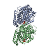
| Structure viewer | Molecule: MolmilJmol/JSmol |
|---|
- Downloads & links
-Download
| PDBx/mmCIF format | 3ono.cif.gz | 98.8 KB | Display | PDBx/mmCIF format |
|---|---|---|---|---|
| PDB format | pdb3ono.ent.gz | 81 KB | Display | PDB format |
| PDBx/mmJSON format | 3ono.json.gz | Tree view | PDBx/mmJSON format | |
| Others |  Other downloads Other downloads |
-Validation report
| Arichive directory | https://data.pdbj.org/pub/pdb/validation_reports/on/3onoftp://data.pdbj.org/pub/pdb/validation_reports/on/3ono | HTTPS FTP |
|---|
-Related structure data
| Similar structure data | |
|---|---|
| Other databases |

-Links
 PDBj
PDBj- Assembly
| Deposited unit | 
| ||||||||
|---|---|---|---|---|---|---|---|---|---|
| 1 | 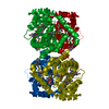
| ||||||||
| Unit cell |
| ||||||||
| Components on special symmetry positions |
|
-Components
| #1: Protein | Mass: 23150.248 Da / Num. of mol.: 1 Source method: isolated from a genetically manipulated source Source: (gene. exp.) Vibrio parahaemolyticus (bacteria) / Strain: RIMD 2210633 / Gene: VPA0080 / Plasmid: pMCSG7 / Production host: Escherichia coli (E. coli) / Strain (production host): BL21magic / References: UniProt: Q87K18 |
|---|---|
| #2: Chemical | ChemComp-GOL / Glycerol  Mass: 92.094 Da / Num. of mol.: 1 / Source method: obtained synthetically / Formula: C3H8O3 Mass: 92.094 Da / Num. of mol.: 1 / Source method: obtained synthetically / Formula: C3H8O3 |
| #3: Water | ChemComp-HOH / Water Mass: 18.015 Da / Num. of mol.: 185 / Source method: isolated from a natural source / Formula: H2O Mass: 18.015 Da / Num. of mol.: 185 / Source method: isolated from a natural source / Formula: H2O |
-Experimental details
-Experiment
| Experiment | Method: X-RAY DIFFRACTION / Number of used crystals: 1 |
|---|
- Sample preparation
| Crystal | Density Matthews: 2.59 Å3/Da / Density % sol: 52.51 % |
|---|---|
| Crystal grow | Temperature: 277 K / Method: vapor diffusion, sitting drop / pH: 7.5 Details: 0.2 M postassium formate, 20 % (v/v) PEG 3350, pH 7.5, VAPOR DIFFUSION, SITTING DROP, temperature 277K |
-Data collection
| Diffraction | Mean temperature: 100 K |
|---|---|
| Diffraction source | Source: SYNCHROTRON / Site: APS  / Beamline: 19-BM / Wavelength: 0.97896 Å / Beamline: 19-BM / Wavelength: 0.97896 Å |
| Detector | Type: SBC-3 / Detector: CCD / Date: Jun 1, 2007 / Details: morrors |
| Radiation | Monochromator: double crystal monochromator / Protocol: SINGLE WAVELENGTH / Monochromatic (M) / Laue (L): M / Scattering type: x-ray |
| Radiation wavelength | Wavelength: 0.97896 Å / Relative weight: 1 |
| Reflection | Resolution: 1.75→50 Å / Num. all: 25080 / Num. obs: 25080 / % possible obs: 98.3 % / Observed criterion σ(F): 0 / Observed criterion σ(I): 0 / Redundancy: 29.1 % / Biso Wilson estimate: 21.74 Å2 / Rsym value: 0.091 / Net I/σ(I): 7.1 |
| Reflection shell | Resolution: 1.75→1.81 Å / Redundancy: 23.9 % / Mean I/σ(I) obs: 4.9 / Num. unique all: 2153 / Rsym value: 0.492 / % possible all: 86.6 |
- Processing
| Software |
| |||||||||||||||||||||||||||||||||||||||||||||||||||||||||||||||||||||||||||||
|---|---|---|---|---|---|---|---|---|---|---|---|---|---|---|---|---|---|---|---|---|---|---|---|---|---|---|---|---|---|---|---|---|---|---|---|---|---|---|---|---|---|---|---|---|---|---|---|---|---|---|---|---|---|---|---|---|---|---|---|---|---|---|---|---|---|---|---|---|---|---|---|---|---|---|---|---|---|---|
| Refinement | Method to determine structure: SAD / Resolution: 1.75→30.452 Å / SU ML: 0.2 / Isotropic thermal model: mixed / Cross valid method: THROUGHOUT / σ(F): 0 / Stereochemistry target values: MLHL
| |||||||||||||||||||||||||||||||||||||||||||||||||||||||||||||||||||||||||||||
| Solvent computation | Shrinkage radii: 1.06 Å / VDW probe radii: 1.3 Å / Solvent model: FLAT BULK SOLVENT MODEL / Bsol: 37.833 Å2 / ksol: 0.337 e/Å3 | |||||||||||||||||||||||||||||||||||||||||||||||||||||||||||||||||||||||||||||
| Displacement parameters | Biso mean: 30.3 Å2
| |||||||||||||||||||||||||||||||||||||||||||||||||||||||||||||||||||||||||||||
| Refinement step | Cycle: LAST / Resolution: 1.75→30.452 Å
| |||||||||||||||||||||||||||||||||||||||||||||||||||||||||||||||||||||||||||||
| Refine LS restraints |
| |||||||||||||||||||||||||||||||||||||||||||||||||||||||||||||||||||||||||||||
| LS refinement shell | Refine-ID: X-RAY DIFFRACTION
| |||||||||||||||||||||||||||||||||||||||||||||||||||||||||||||||||||||||||||||
| Refinement TLS params. | Method: refined / Origin x: 11.2483 Å / Origin y: 3.3129 Å / Origin z: 18.9137 Å
| |||||||||||||||||||||||||||||||||||||||||||||||||||||||||||||||||||||||||||||
| Refinement TLS group | Selection details: chain A |
