 Movie
Movie Controller

[English] 日本語
 Yorodumi
- PDB-3oml: Structure of full-length peroxisomal multifunctional enzyme type ... -
+ Open data

- Basic information
| Entry | Database: PDB / ID: 3oml | ||||||
|---|---|---|---|---|---|---|---|
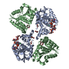
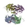
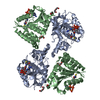
| Title | Structure of full-length peroxisomal multifunctional enzyme type 2 from Drosophila melanogaster | ||||||
 Components Components | Peroxisomal Multifunctional Enzyme Type 2, CG3415 | ||||||
 Keywords Keywords |  Oxidoreductase / Hydrolase / Rossmann fold / hot-dog fold / hydratase 2 motif / Peroxisomes / lyase Oxidoreductase / Hydrolase / Rossmann fold / hot-dog fold / hydratase 2 motif / Peroxisomes / lyase | ||||||
| Function / homology |  Function and homology information Function and homology informationSynthesis of bile acids and bile salts via 7alpha-hydroxycholesterol / alpha-linolenic acid (ALA) metabolism / Beta-oxidation of pristanoyl-CoA / Beta-oxidation of very long chain fatty acids / Peroxisomal protein import / (3R)-hydroxyacyl-CoA dehydrogenase (NAD+) activity / : / enoyl-CoA hydratase 2 / 3-hydroxyacyl-CoA dehydratase activity / fatty acid beta-oxidation using acyl-CoA oxidase ...Synthesis of bile acids and bile salts via 7alpha-hydroxycholesterol / alpha-linolenic acid (ALA) metabolism / Beta-oxidation of pristanoyl-CoA / Beta-oxidation of very long chain fatty acids / Peroxisomal protein import / (3R)-hydroxyacyl-CoA dehydrogenase (NAD+) activity / : / enoyl-CoA hydratase 2 / 3-hydroxyacyl-CoA dehydratase activity / fatty acid beta-oxidation using acyl-CoA oxidase / 17-beta-hydroxysteroid dehydrogenase (NAD+) activity / 3-hydroxyacyl-CoA dehydrogenase activity / estradiol 17-beta-dehydrogenase [NAD(P)] activity / enoyl-CoA hydratase activity / fatty acid beta-oxidation / peroxisome / protein homodimerization activitySimilarity search - Function | ||||||
| Biological species |  Drosophila melanogaster (fruit fly) Drosophila melanogaster (fruit fly) | ||||||
| Method | X-RAY DIFFRACTION / SYNCHROTRON / MOLECULAR REPLACEMENT / Resolution: 2.15 Å | ||||||
 Authors Authors | Haataja, T.J.K. / Koski, M.K. / Glumoff, T. / Hiltunen, J.K. | ||||||
 Citation Citation | Journal: Biochem.J. / Year: 2011 Title: Peroxisomal multifunctional enzyme type 2 from the fruitfly: dehydrogenase and hydratase act as separate entities, as revealed by structure and kinetics. Authors: Haataja, T.J. / Koski, M.K. / Hiltunen, J.K. / Glumoff, T. | ||||||
| History |
|
- Structure visualization
| Structure viewer | Molecule: MolmilJmol/JSmol |
|---|
- Downloads & links
-Download
| PDBx/mmCIF format | 3oml.cif.gz | 225.4 KB | Display | PDBx/mmCIF format |
|---|---|---|---|---|
| PDB format | pdb3oml.ent.gz | 179.9 KB | Display | PDB format |
| PDBx/mmJSON format | 3oml.json.gz | Tree view | PDBx/mmJSON format | |
| Others |  Other downloads Other downloads |
-Validation report
| Arichive directory | https://data.pdbj.org/pub/pdb/validation_reports/om/3omlftp://data.pdbj.org/pub/pdb/validation_reports/om/3oml | HTTPS FTP |
|---|
-Related structure data


-Links
 PDBj
PDBj


- Assembly
| Deposited unit | 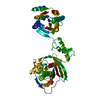
| ||||||||
|---|---|---|---|---|---|---|---|---|---|
| 1 | 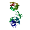
| ||||||||
| Unit cell |
|
-Components
| #1: Protein | Mass: 66174.148 Da / Num. of mol.: 1 Source method: isolated from a genetically manipulated source Source: (gene. exp.) Drosophila melanogaster (fruit fly) / Gene: CG3415, Dmel_CG3415 / Production host:  Escherichia coli (E. coli) / Strain (production host): BL21(DE3)pLysS Escherichia coli (E. coli) / Strain (production host): BL21(DE3)pLysSReferences: UniProt: Q9VXJ0, 17beta-estradiol 17-dehydrogenase, Hydrolases; Acting on peptide bonds (peptidases); Cysteine endopeptidases |
|---|---|
| #2: Water | ChemComp-HOH / Water Mass: 18.015 Da / Num. of mol.: 138 / Source method: isolated from a natural source / Formula: H2O Mass: 18.015 Da / Num. of mol.: 138 / Source method: isolated from a natural source / Formula: H2O |
-Experimental details
-Experiment
| Experiment | Method: X-RAY DIFFRACTION / Number of used crystals: 1 |
|---|
- Sample preparation
| Crystal | Density Matthews: 2.21 Å3/Da / Density % sol: 44.24 % |
|---|---|
| Crystal grow | Temperature: 294 K / Method: vapor diffusion, sitting drop / pH: 8 Details: 0.1 M Tris, 1.0 M NaCl, 20 % (w/v) PEG 5000 MME, 5 mM NAD+ , VAPOR DIFFUSION, SITTING DROP, temperature 294K, pH 8.0 |
-Data collection
| Diffraction | Mean temperature: 100 K |
|---|---|
| Diffraction source | Source: SYNCHROTRON / Site: ESRF  / Beamline: ID29 / Wavelength: 0.931 Å / Beamline: ID29 / Wavelength: 0.931 Å |
| Detector | Type: ADSC QUANTUM 315r / Detector: CCD / Date: Feb 25, 2008 |
| Radiation | Monochromator: Si 311 CHANNEL / Protocol: SINGLE WAVELENGTH / Monochromatic (M) / Laue (L): M / Scattering type: x-ray |
| Radiation wavelength | Wavelength: 0.931 Å / Relative weight: 1 |
| Reflection | Resolution: 2.15→28.1 Å / Num. all: 31114 / Num. obs: 31008 / % possible obs: 99.66 % |
| Reflection shell | Resolution: 2.15→2.21 Å / % possible all: 99.7 |
- Processing
| Software |
| ||||||||||||||||||||||||||||||||||||||||||||||||||||||||||||||||||||||||||||||||||||||||||||||||||||||||||||||||||||||||||||||||||||||||||||||||||||||||||||||||||||||||||
|---|---|---|---|---|---|---|---|---|---|---|---|---|---|---|---|---|---|---|---|---|---|---|---|---|---|---|---|---|---|---|---|---|---|---|---|---|---|---|---|---|---|---|---|---|---|---|---|---|---|---|---|---|---|---|---|---|---|---|---|---|---|---|---|---|---|---|---|---|---|---|---|---|---|---|---|---|---|---|---|---|---|---|---|---|---|---|---|---|---|---|---|---|---|---|---|---|---|---|---|---|---|---|---|---|---|---|---|---|---|---|---|---|---|---|---|---|---|---|---|---|---|---|---|---|---|---|---|---|---|---|---|---|---|---|---|---|---|---|---|---|---|---|---|---|---|---|---|---|---|---|---|---|---|---|---|---|---|---|---|---|---|---|---|---|---|---|---|---|---|---|---|
| Refinement | Method to determine structure: MOLECULAR REPLACEMENT Starting model: PDB ENTRIES 1PN4 AND 1GZ6 Resolution: 2.15→28.1 Å / Cor.coef. Fo:Fc: 0.941 / Cor.coef. Fo:Fc free: 0.914 / SU B: 18.872 / SU ML: 0.212 / Cross valid method: THROUGHOUT / ESU R: 0.268 / ESU R Free: 0.226 / Stereochemistry target values: MAXIMUM LIKELIHOOD / Details: HYDROGENS HAVE BEEN ADDED IN THE RIDING POSITIONS
| ||||||||||||||||||||||||||||||||||||||||||||||||||||||||||||||||||||||||||||||||||||||||||||||||||||||||||||||||||||||||||||||||||||||||||||||||||||||||||||||||||||||||||
| Solvent computation | Ion probe radii: 0.8 Å / Shrinkage radii: 0.8 Å / VDW probe radii: 1.4 Å / Solvent model: MASK | ||||||||||||||||||||||||||||||||||||||||||||||||||||||||||||||||||||||||||||||||||||||||||||||||||||||||||||||||||||||||||||||||||||||||||||||||||||||||||||||||||||||||||
| Displacement parameters | Biso mean: 65.23 Å2
| ||||||||||||||||||||||||||||||||||||||||||||||||||||||||||||||||||||||||||||||||||||||||||||||||||||||||||||||||||||||||||||||||||||||||||||||||||||||||||||||||||||||||||
| Refinement step | Cycle: LAST / Resolution: 2.15→28.1 Å
| ||||||||||||||||||||||||||||||||||||||||||||||||||||||||||||||||||||||||||||||||||||||||||||||||||||||||||||||||||||||||||||||||||||||||||||||||||||||||||||||||||||||||||
| Refine LS restraints |
| ||||||||||||||||||||||||||||||||||||||||||||||||||||||||||||||||||||||||||||||||||||||||||||||||||||||||||||||||||||||||||||||||||||||||||||||||||||||||||||||||||||||||||
| LS refinement shell | Resolution: 2.15→2.205 Å / Total num. of bins used: 20
| ||||||||||||||||||||||||||||||||||||||||||||||||||||||||||||||||||||||||||||||||||||||||||||||||||||||||||||||||||||||||||||||||||||||||||||||||||||||||||||||||||||||||||
| Refinement TLS params. | Method: refined / Refine-ID: X-RAY DIFFRACTION
| ||||||||||||||||||||||||||||||||||||||||||||||||||||||||||||||||||||||||||||||||||||||||||||||||||||||||||||||||||||||||||||||||||||||||||||||||||||||||||||||||||||||||||
| Refinement TLS group |
|
