 Movie
Movie Controller


+ Open data

- Basic information
| Entry | Database: PDB / ID: 3oit | ||||||
|---|---|---|---|---|---|---|---|
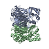
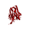
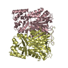
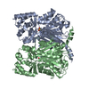
| Title | Crystal structure of curcuminoid synthase CUS from Oryza sativa | ||||||
 Components Components | Os07g0271500 protein | ||||||
 Keywords Keywords |  TRANSFERASE / type III polyketide synthases TRANSFERASE / type III polyketide synthases | ||||||
| Function / homology |  Function and homology information Function and homology informationbisdemethoxycurcumin synthase / bisdemethoxycurcumin synthase activity / flavonoid biosynthetic process / polyketide biosynthetic process / acyltransferase activity, transferring groups other than amino-acyl groups / identical protein binding Similarity search - Function | ||||||
| Biological species |  Oryza sativa (Asian cultivated rice) Oryza sativa (Asian cultivated rice) | ||||||
| Method | X-RAY DIFFRACTION / SYNCHROTRON / MOLECULAR REPLACEMENT / Resolution: 2 Å | ||||||
 Authors Authors | Miyazono, K. / Um, J. / Imai, F.L. / Katsuyama, Y. / Ohnishi, Y. / Horinouchi, S. / Tanokura, M. | ||||||
 Citation Citation | Journal: Proteins / Year: 2011 Title: Crystal structure of curcuminoid synthase CUS from Oryza sativa Authors: Miyazono, K. / Um, J. / Imai, F.L. / Katsuyama, Y. / Ohnishi, Y. / Horinouchi, S. / Tanokura, M. | ||||||
| History |
|
- Structure visualization
| Structure viewer | Molecule: MolmilJmol/JSmol |
|---|
- Downloads & links
-Download
| PDBx/mmCIF format | 3oit.cif.gz | 292.6 KB | Display | PDBx/mmCIF format |
|---|---|---|---|---|
| PDB format | pdb3oit.ent.gz | 236.5 KB | Display | PDB format |
| PDBx/mmJSON format | 3oit.json.gz | Tree view | PDBx/mmJSON format | |
| Others |  Other downloads Other downloads |
-Validation report
| Arichive directory | https://data.pdbj.org/pub/pdb/validation_reports/oi/3oitftp://data.pdbj.org/pub/pdb/validation_reports/oi/3oit | HTTPS FTP |
|---|
-Related structure data
| Related structure data | 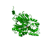 1bi5S S: Starting model for refinement |
|---|---|
| Similar structure data |


-Links
 PDBj
PDBj

- Assembly
| Deposited unit | 
| ||||||||
|---|---|---|---|---|---|---|---|---|---|
| 1 |
| ||||||||
| Unit cell |
|
-Components
| #1: Protein | Mass: 41805.637 Da / Num. of mol.: 2 / Fragment: UNP residues 17-400 Source method: isolated from a genetically manipulated source Source: (gene. exp.) Oryza sativa (Asian cultivated rice) / Strain: subsp. japonica / Gene: OJ1001_C01.122, OSJNBb0002J01.6, Os07g0271500 / Plasmid: pET26b / Production host:  Escherichia coli (E. coli) / References: UniProt: Q8LIL0 Escherichia coli (E. coli) / References: UniProt: Q8LIL0#2: Water | ChemComp-HOH / | Water Mass: 18.015 Da / Num. of mol.: 438 / Source method: isolated from a natural source / Formula: H2O Mass: 18.015 Da / Num. of mol.: 438 / Source method: isolated from a natural source / Formula: H2OSequence details | CDNA WITH ACCESSION NUMBER AK109558 WAS USED FOR THE PRODUCTION OF PROTEIN CUS. AND AUTHOR STATED ...CDNA WITH ACCESSION NUMBER AK109558 WAS USED FOR THE PRODUCTION | |
|---|
-Experimental details
-Experiment
| Experiment | Method: X-RAY DIFFRACTION / Number of used crystals: 1 |
|---|
- Sample preparation
| Crystal | Density Matthews: 2.51 Å3/Da / Density % sol: 50.92 % |
|---|---|
| Crystal grow | Temperature: 293 K / Method: vapor diffusion, sitting drop / pH: 8.5 Details: 100mM Tris-HCl, 20% PEG 4000, 200mM NaCl, 6% 1,6-hexanediol, pH 8.5, VAPOR DIFFUSION, SITTING DROP, temperature 293K |
-Data collection
| Diffraction | Mean temperature: 95 K | |||||||||||||||||||||||||||||||||||||||||||||||||||||||||||||||||||||||||||||||||||||||||||||||||||||||||||||||||||||||||||||||||||||||||||||||||||||||||||||||||||||||||||||||||||||||||||||
|---|---|---|---|---|---|---|---|---|---|---|---|---|---|---|---|---|---|---|---|---|---|---|---|---|---|---|---|---|---|---|---|---|---|---|---|---|---|---|---|---|---|---|---|---|---|---|---|---|---|---|---|---|---|---|---|---|---|---|---|---|---|---|---|---|---|---|---|---|---|---|---|---|---|---|---|---|---|---|---|---|---|---|---|---|---|---|---|---|---|---|---|---|---|---|---|---|---|---|---|---|---|---|---|---|---|---|---|---|---|---|---|---|---|---|---|---|---|---|---|---|---|---|---|---|---|---|---|---|---|---|---|---|---|---|---|---|---|---|---|---|---|---|---|---|---|---|---|---|---|---|---|---|---|---|---|---|---|---|---|---|---|---|---|---|---|---|---|---|---|---|---|---|---|---|---|---|---|---|---|---|---|---|---|---|---|---|---|---|---|---|
| Diffraction source | Source: SYNCHROTRON / Site: Photon Factory  / Beamline: AR-NW12A / Wavelength: 1 Å / Beamline: AR-NW12A / Wavelength: 1 Å | |||||||||||||||||||||||||||||||||||||||||||||||||||||||||||||||||||||||||||||||||||||||||||||||||||||||||||||||||||||||||||||||||||||||||||||||||||||||||||||||||||||||||||||||||||||||||||||
| Detector | Type: ADSC QUANTUM 210r / Detector: CCD / Date: Feb 7, 2010 | |||||||||||||||||||||||||||||||||||||||||||||||||||||||||||||||||||||||||||||||||||||||||||||||||||||||||||||||||||||||||||||||||||||||||||||||||||||||||||||||||||||||||||||||||||||||||||||
| Radiation | Protocol: SINGLE WAVELENGTH / Monochromatic (M) / Laue (L): M / Scattering type: x-ray | |||||||||||||||||||||||||||||||||||||||||||||||||||||||||||||||||||||||||||||||||||||||||||||||||||||||||||||||||||||||||||||||||||||||||||||||||||||||||||||||||||||||||||||||||||||||||||||
| Radiation wavelength | Wavelength: 1 Å / Relative weight: 1 | |||||||||||||||||||||||||||||||||||||||||||||||||||||||||||||||||||||||||||||||||||||||||||||||||||||||||||||||||||||||||||||||||||||||||||||||||||||||||||||||||||||||||||||||||||||||||||||
| Reflection | Resolution: 2→20 Å / Num. obs: 57647 / % possible obs: 99.7 % / Observed criterion σ(I): -3 / Biso Wilson estimate: 38.965 Å2 / Rmerge(I) obs: 0.065 / Net I/σ(I): 22.48 | |||||||||||||||||||||||||||||||||||||||||||||||||||||||||||||||||||||||||||||||||||||||||||||||||||||||||||||||||||||||||||||||||||||||||||||||||||||||||||||||||||||||||||||||||||||||||||||
| Reflection shell | Diffraction-ID: 1
|
- Processing
| Software |
| |||||||||||||||||||||||||||||||||||||||||||||||||||||||||||||||||||||||||||
|---|---|---|---|---|---|---|---|---|---|---|---|---|---|---|---|---|---|---|---|---|---|---|---|---|---|---|---|---|---|---|---|---|---|---|---|---|---|---|---|---|---|---|---|---|---|---|---|---|---|---|---|---|---|---|---|---|---|---|---|---|---|---|---|---|---|---|---|---|---|---|---|---|---|---|---|---|
| Refinement | Method to determine structure: MOLECULAR REPLACEMENT Starting model: PDB ENTRY 1BI5 Resolution: 2→20 Å / Cor.coef. Fo:Fc: 0.959 / Cor.coef. Fo:Fc free: 0.946 / Occupancy max: 1 / Occupancy min: 1 / SU B: 8.808 / SU ML: 0.109 / Cross valid method: THROUGHOUT / σ(F): 0 / ESU R Free: 0.152 / Stereochemistry target values: MAXIMUM LIKELIHOOD
| |||||||||||||||||||||||||||||||||||||||||||||||||||||||||||||||||||||||||||
| Solvent computation | Ion probe radii: 0.8 Å / Shrinkage radii: 0.8 Å / VDW probe radii: 1.4 Å / Solvent model: MASK | |||||||||||||||||||||||||||||||||||||||||||||||||||||||||||||||||||||||||||
| Displacement parameters | Biso max: 60.16 Å2 / Biso mean: 36.755 Å2 / Biso min: 16.43 Å2
| |||||||||||||||||||||||||||||||||||||||||||||||||||||||||||||||||||||||||||
| Refinement step | Cycle: LAST / Resolution: 2→20 Å
| |||||||||||||||||||||||||||||||||||||||||||||||||||||||||||||||||||||||||||
| Refine LS restraints |
| |||||||||||||||||||||||||||||||||||||||||||||||||||||||||||||||||||||||||||
| LS refinement shell | Resolution: 2→2.052 Å / Total num. of bins used: 20
| |||||||||||||||||||||||||||||||||||||||||||||||||||||||||||||||||||||||||||
| Refinement TLS params. | Method: refined / Refine-ID: X-RAY DIFFRACTION
| |||||||||||||||||||||||||||||||||||||||||||||||||||||||||||||||||||||||||||
| Refinement TLS group |
|
