 Movie
Movie Controller

[English] 日本語
 Yorodumi
- PDB-3oi8: The crystal structure of functionally unknown conserved protein d... -
+ Open data

- Basic information
| Entry | Database: PDB / ID: 3oi8 | ||||||
|---|---|---|---|---|---|---|---|
| Title | The crystal structure of functionally unknown conserved protein domain from Neisseria meningitidis MC58 | ||||||
 Components Components | Uncharacterized protein | ||||||
 Keywords Keywords |  Structural genomics / Unknown function / PSI-2 / protein structure initiative / midwest center for structural genomics / MCSG Structural genomics / Unknown function / PSI-2 / protein structure initiative / midwest center for structural genomics / MCSG | ||||||
| Function / homology |  Function and homology informationflavin adenine dinucleotide binding / metal ion binding / plasma membrane Function and homology informationflavin adenine dinucleotide binding / metal ion binding / plasma membraneSimilarity search - Function | ||||||
| Biological species |  Neisseria meningitidis serogroup B (bacteria) Neisseria meningitidis serogroup B (bacteria) | ||||||
| Method | X-RAY DIFFRACTION / SYNCHROTRON / MAD / Resolution: 1.989 Å | ||||||
 Authors Authors | Zhang, R. / Tan, K. / Li, H. / Cobb, G. / Joachimiak, A. / Midwest Center for Structural Genomics (MCSG) | ||||||
 Citation Citation | Journal: To be Published Title: The crystal structure of functionally unknown conserved protein domain from Neisseria meningitidis MC58 Authors: Zhang, R. / Tan, K. / Li, H. / Cobb, G. / Joachimiak, A. | ||||||
| History |
|
- Structure visualization
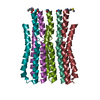
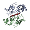
| Structure viewer | Molecule: MolmilJmol/JSmol |
|---|
- Downloads & links
-Download
| PDBx/mmCIF format | 3oi8.cif.gz | 136.9 KB | Display | PDBx/mmCIF format |
|---|---|---|---|---|
| PDB format | pdb3oi8.ent.gz | 114.2 KB | Display | PDB format |
| PDBx/mmJSON format | 3oi8.json.gz | Tree view | PDBx/mmJSON format | |
| Others |  Other downloads Other downloads |
-Validation report
| Arichive directory | https://data.pdbj.org/pub/pdb/validation_reports/oi/3oi8ftp://data.pdbj.org/pub/pdb/validation_reports/oi/3oi8 | HTTPS FTP |
|---|
-Related structure data
| Similar structure data | |
|---|---|
| Other databases |


-Links
 PDBj
PDBj


- Assembly
| Deposited unit | 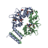
| ||||||||
|---|---|---|---|---|---|---|---|---|---|
| 1 |
| ||||||||
| Unit cell |
| ||||||||
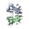
| Details | Experimentally unknown. It is predicted that the chains A and B form a dimer. |
-Components
| #1: Protein | Mass: 18040.861 Da / Num. of mol.: 2 / Fragment: sequence database residues 24-176 Source method: isolated from a genetically manipulated source Source: (gene. exp.) Neisseria meningitidis serogroup B (bacteria)Strain: MC58 / Gene: NMB0537 / Plasmid: pMCSG19 / Production host: Escherichia coli (E. coli) / Strain (production host): pPK1037 / References: UniProt: Q9K0P8#2: Chemical | Adenosine  Mass: 267.241 Da / Num. of mol.: 2 / Source method: obtained synthetically / Formula: C10H13N5O4 Mass: 267.241 Da / Num. of mol.: 2 / Source method: obtained synthetically / Formula: C10H13N5O4#3: Chemical | ChemComp-GOL / | Glycerol  Mass: 92.094 Da / Num. of mol.: 1 / Source method: obtained synthetically / Formula: C3H8O3 Mass: 92.094 Da / Num. of mol.: 1 / Source method: obtained synthetically / Formula: C3H8O3#4: Chemical | ChemComp-PEG / | Diethylene glycol  Mass: 106.120 Da / Num. of mol.: 1 / Source method: obtained synthetically / Formula: C4H10O3 Mass: 106.120 Da / Num. of mol.: 1 / Source method: obtained synthetically / Formula: C4H10O3#5: Water | ChemComp-HOH / | Water Mass: 18.015 Da / Num. of mol.: 142 / Source method: isolated from a natural source / Formula: H2O Mass: 18.015 Da / Num. of mol.: 142 / Source method: isolated from a natural source / Formula: H2O |
|---|
-Experimental details
-Experiment
| Experiment | Method: X-RAY DIFFRACTION / Number of used crystals: 1 |
|---|
- Sample preparation
| Crystal | Density Matthews: 2.29 Å3/Da / Density % sol: 46.28 % |
|---|---|
| Crystal grow | Temperature: 289 K / Method: vapor diffusion, sitting drop / pH: 8.5 Details: 20% w/v PEG8000, 0.1M Tris, 0.2M MgCl2, pH 8.5, VAPOR DIFFUSION, SITTING DROP, temperature 289K |
-Data collection
| Diffraction | Mean temperature: 100 K | |||||||||
|---|---|---|---|---|---|---|---|---|---|---|
| Diffraction source | Source: SYNCHROTRON / Site: APS  / Beamline: 19-ID / Wavelength: 0.97926, 0.97942 / Beamline: 19-ID / Wavelength: 0.97926, 0.97942 | |||||||||
| Detector | Type: ADSC QUANTUM 315r / Detector: CCD / Date: Jul 2, 2010 / Details: mirror | |||||||||
| Radiation | Monochromator: Si 111 crystal / Protocol: MAD / Monochromatic (M) / Laue (L): M / Scattering type: x-ray | |||||||||
| Radiation wavelength |
| |||||||||
| Reflection | Resolution: 2→39.1 Å / Num. all: 22323 / Num. obs: 22323 / % possible obs: 98.4 % / Observed criterion σ(F): 0 / Observed criterion σ(I): 0 / Redundancy: 4.7 % / Biso Wilson estimate: 25.97 Å2 / Rmerge(I) obs: 0.134 / Net I/σ(I): 16.2 | |||||||||
| Reflection shell | Resolution: 2→2.03 Å / Redundancy: 3.1 % / Rmerge(I) obs: 0.52 / Mean I/σ(I) obs: 1.07 / % possible all: 72 |
- Processing
| Software |
| |||||||||||||||||||||||||||||||||||||||||||||||||||||||||||||||||||||||||||
|---|---|---|---|---|---|---|---|---|---|---|---|---|---|---|---|---|---|---|---|---|---|---|---|---|---|---|---|---|---|---|---|---|---|---|---|---|---|---|---|---|---|---|---|---|---|---|---|---|---|---|---|---|---|---|---|---|---|---|---|---|---|---|---|---|---|---|---|---|---|---|---|---|---|---|---|---|
| Refinement | Method to determine structure: MAD / Resolution: 1.989→39.095 Å / SU ML: 0.23 / σ(F): 0.03 / Phase error: 25.09 / Stereochemistry target values: ML
| |||||||||||||||||||||||||||||||||||||||||||||||||||||||||||||||||||||||||||
| Solvent computation | Shrinkage radii: 0.9 Å / VDW probe radii: 1.11 Å / Solvent model: FLAT BULK SOLVENT MODEL / Bsol: 39.3 Å2 / ksol: 0.373 e/Å3 | |||||||||||||||||||||||||||||||||||||||||||||||||||||||||||||||||||||||||||
| Displacement parameters |
| |||||||||||||||||||||||||||||||||||||||||||||||||||||||||||||||||||||||||||
| Refinement step | Cycle: LAST / Resolution: 1.989→39.095 Å
| |||||||||||||||||||||||||||||||||||||||||||||||||||||||||||||||||||||||||||
| Refine LS restraints |
| |||||||||||||||||||||||||||||||||||||||||||||||||||||||||||||||||||||||||||
| LS refinement shell |
| |||||||||||||||||||||||||||||||||||||||||||||||||||||||||||||||||||||||||||
| Refinement TLS params. | Method: refined / Refine-ID: X-RAY DIFFRACTION
| |||||||||||||||||||||||||||||||||||||||||||||||||||||||||||||||||||||||||||
| Refinement TLS group |
|
