Journal: To be Published Title: Non-Resonance Raman Difference Spectroscopy as a Tool to Probe Enthalpy-Entropy Compensation and the Interfacial Mobility Model Authors: Kowatz, T. / Naismith, J.H.
Resolution: 2.5→25.6 Å / Cor.coef. Fo:Fc: 0.909 / Cor.coef. Fo:Fc free: 0.885 / SU B: 24.34 / SU ML: 0.266 / Cross valid method: THROUGHOUT / ESU R Free: 0.328 / Stereochemistry target values: MAXIMUM LIKELIHOOD / Details: HYDROGENS HAVE BEEN USED IF PRESENT IN THE INPUT
Rfactor
Num. reflection
% reflection
Selection details
Rfree
0.26448
276
4.7 %
RANDOM
Rwork
0.25466
-
-
-
obs
0.25512
5540
99.03 %
-
Solvent computation
Ion probe radii: 0.8 Å / Shrinkage radii: 0.8 Å / VDW probe radii: 1.4 Å / Solvent model: BABINET MODEL WITH MASK
Displacement parameters
Biso mean: 94.18 Å2
Baniso -1
Baniso -2
Baniso -3
1-
3.73 Å2
-0 Å2
0 Å2
2-
-
-2.12 Å2
0 Å2
3-
-
-
-1.61 Å2
Refinement step
Cycle: LAST / Resolution: 2.5→25.6 Å
Protein
Nucleic acid
Ligand
Solvent
Total
Num. atoms
1271
0
27
15
1313
Refine LS restraints
Refine-ID
Type
Dev ideal
Dev ideal target
Number
X-RAY DIFFRACTION
r_bond_refined_d
0.007
0.022
1356
X-RAY DIFFRACTION
r_bond_other_d
0.001
0.02
892
X-RAY DIFFRACTION
r_angle_refined_deg
0.782
1.957
1818
X-RAY DIFFRACTION
r_angle_other_deg
0.757
3.002
2144
X-RAY DIFFRACTION
r_dihedral_angle_1_deg
5.205
5
156
X-RAY DIFFRACTION
r_dihedral_angle_2_deg
30.024
23.485
66
X-RAY DIFFRACTION
r_dihedral_angle_3_deg
10.441
15
190
X-RAY DIFFRACTION
r_dihedral_angle_4_deg
7.519
15
7
X-RAY DIFFRACTION
r_chiral_restr
0.046
0.2
187
X-RAY DIFFRACTION
r_gen_planes_refined
0.003
0.021
1488
X-RAY DIFFRACTION
r_gen_planes_other
0.001
0.02
292
LS refinement shell
Resolution: 2.498→2.563 Å / Total num. of bins used: 20
Rfactor
Num. reflection
% reflection
Rfree
0.212
22
-
Rwork
0.262
354
-
obs
-
-
90.6 %
Refinement TLS params.
Method: refined / Refine-ID: X-RAY DIFFRACTION
ID
L11 (°2)
L12 (°2)
L13 (°2)
L22 (°2)
L23 (°2)
L33 (°2)
S11 (Å °)
S12 (Å °)
S13 (Å °)
S21 (Å °)
S22 (Å °)
S23 (Å °)
S31 (Å °)
S32 (Å °)
S33 (Å °)
T11 (Å2)
T12 (Å2)
T13 (Å2)
T22 (Å2)
T23 (Å2)
T33 (Å2)
Origin x (Å)
Origin y (Å)
Origin z (Å)
1
2.3852
0.5243
-0.3692
9.9282
1.1924
3.2868
0.2031
-0.1739
-0.0649
1.5716
-0.1389
-1.0764
0.3701
0.3706
-0.0642
0.4342
-0.0174
-0.1454
0.405
0.0996
0.4187
8.683
-15.266
1.108
2
2.5982
-0.4037
-0.3707
6.3368
1.7659
5.7849
0.0167
-0.1208
-0.169
0.5718
-0.0015
-0.698
0.3648
0.2936
-0.0152
0.2887
0.0037
-0.0347
0.3697
0.04
0.3627
8.738
-14.999
-4.487
3
1.9609
-3.297
-2.5286
9.8252
-0.2051
8.5845
-0.1541
-0.2605
0.7185
0.9337
0.1131
-2.5348
-0.0351
0.8422
0.041
0.5124
0.0947
-0.3947
0.5392
0.0316
1.1932
18.895
-14.326
-2.253
4
23.2106
2.1765
-6.4669
7.4055
-4.4867
3.9054
-0.5568
-1.6977
3.3787
1.0469
-0.6418
-3.5383
-0.3796
0.8696
1.1986
2.3315
0.8299
-1.9258
1.2547
-0.5883
3.9532
23.424
-17.38
4.52
5
0.3273
0.4248
-0.0626
0.5613
-0.0972
0.2067
0.0305
0.0389
0.2496
0.045
0.0149
0.3967
-0.1673
0.1356
-0.0454
0.62
0.2384
0.1843
0.4081
-0.0127
0.9156
8.17
-19.563
-17.109
6
0.11
-0.174
0.0947
0.3143
-0.1865
0.1473
-0.0216
0.0001
0.0856
0.0764
-0.0611
-0.1771
0.0453
0.114
0.0827
0.502
0.0497
-0.069
0.3785
0.0154
0.2495
3.223
-9.07
2.317
7
0.1574
0.0961
0.0432
0.0788
0.0552
0.0613
-0.0084
0.012
0.1869
-0.0527
0
0.0691
-0.0634
0.0058
0.0084
0.3071
-0.0499
0.0876
0.3663
0.0206
0.3958
13.443
-11.305
-16.684
8
0.0268
-0.0242
0.0029
0.023
0.0004
0.0089
-0.0011
0.003
0.0571
-0.0068
-0.0065
-0.0475
-0.0271
-0.0079
0.0076
0.336
-0.0234
-0.0292
0.3425
0.056
0.3869
6.808
-7.637
-8.573
9
0.0608
0.0668
0.0144
0.1988
-0.0003
0.0418
-0.0004
-0.0407
0.0436
-0.0446
0.0257
0.154
-0.0099
0.1033
-0.0252
0.315
-0.0294
-0.059
0.5317
0.1379
0.8608
18.298
-9.999
-4.926
10
0.5811
0.2247
-6.1119
46.6217
7.6144
66.4786
0.1314
-0.0848
-0.1071
-0.2245
-1.0807
-0.6974
-1.5433
0.7689
0.9493
0.0469
-0.0618
-0.006
0.2403
0.0277
0.2439
18.2906
-11.9922
-8.2683
Refinement TLS group
ID
Refine-ID
Refine TLS-ID
Auth asym-ID
Auth seq-ID
1
X-RAY DIFFRACTION
1
A
83 - 116
2
X-RAY DIFFRACTION
2
A
117 - 192
3
X-RAY DIFFRACTION
3
A
193 - 220
4
X-RAY DIFFRACTION
4
A
221 - 249
5
X-RAY DIFFRACTION
5
A
1
6
X-RAY DIFFRACTION
6
A
2
7
X-RAY DIFFRACTION
7
A
3
8
X-RAY DIFFRACTION
8
A
4
9
X-RAY DIFFRACTION
9
A
5
10
X-RAY DIFFRACTION
10
A
252
+
About Yorodumi
-
News
-
Feb 9, 2022. New format data for meta-information of EMDB entries
New format data for meta-information of EMDB entries
Version 3 of the EMDB header file is now the official format.
The previous official version 1.9 will be removed from the archive.
In the structure databanks used in Yorodumi, some data are registered as the other names, "COVID-19 virus" and "2019-nCoV". Here are the details of the virus and the list of structure data.
Jan 31, 2019. EMDB accession codes are about to change! (news from PDBe EMDB page)
EMDB accession codes are about to change! (news from PDBe EMDB page)
The allocation of 4 digits for EMDB accession codes will soon come to an end. Whilst these codes will remain in use, new EMDB accession codes will include an additional digit and will expand incrementally as the available range of codes is exhausted. The current 4-digit format prefixed with “EMD-” (i.e. EMD-XXXX) will advance to a 5-digit format (i.e. EMD-XXXXX), and so on. It is currently estimated that the 4-digit codes will be depleted around Spring 2019, at which point the 5-digit format will come into force.
The EM Navigator/Yorodumi systems omit the EMD- prefix.
Related info.:Q: What is EMD? / ID/Accession-code notation in Yorodumi/EM Navigator
Yorodumi is a browser for structure data from EMDB, PDB, SASBDB, etc.
This page is also the successor to EM Navigator detail page, and also detail information page/front-end page for Omokage search.
The word "yorodu" (or yorozu) is an old Japanese word meaning "ten thousand". "mi" (miru) is to see.
Related info.:EMDB / PDB / SASBDB / Comparison of 3 databanks / Yorodumi Search / Aug 31, 2016. New EM Navigator & Yorodumi / Yorodumi Papers / Jmol/JSmol / Function and homology information / Changes in new EM Navigator and Yorodumi
 Movie
Movie Controller

 Yorodumi
 Open data
 Basic information
 Components
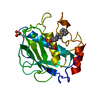
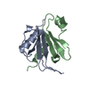
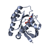
 Matrix metalloproteinase
  Keywords
 Function and homology information
 Authors
 Citation
 Structure visualization
 Downloads & links
 Other downloads


 PDBj
PDBj


 Assembly


 Mass: 40.078 Da / Num. of mol.: 3 / Source method: obtained synthetically / Formula: Ca
 Mass: 65.409 Da / Num. of mol.: 2 / Source method: obtained synthetically / Formula: Zn
 Mass: 260.267 Da / Num. of mol.: 1 / Source method: obtained synthetically / Formula: C9H12N2O5S
 Mass: 96.063 Da / Num. of mol.: 1 / Source method: obtained synthetically / Formula: SO4
 Sample preparation
 Processing