 Movie
Movie Controller

[English] 日本語
 Yorodumi
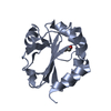
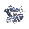
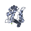
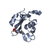
- PDB-3ogn: Crystal Structure of an Odorant-binding Protein from the Southern... -
+ Open data

- Basic information
| Entry | Database: PDB / ID: 3ogn | ||||||
|---|---|---|---|---|---|---|---|
| Title | Crystal Structure of an Odorant-binding Protein from the Southern House Mosquito Complexed with an Oviposition Pheromone | ||||||
 Components Components | Odorant-binding protein | ||||||
 Keywords Keywords | TRANSPORT PROTEIN / helix bundle / Odorant-binding Protein | ||||||
| Function / homology |  Function and homology information Function and homology information | ||||||
| Biological species |  Culex quinquefasciatus (southern house mosquito) Culex quinquefasciatus (southern house mosquito) | ||||||
| Method | X-RAY DIFFRACTION / SYNCHROTRON / MOLECULAR REPLACEMENT / Resolution: 1.3 Å | ||||||
 Authors Authors | Mao, Y. / Clardy, J. | ||||||
 Citation Citation | Journal: Proc.Natl.Acad.Sci.USA / Year: 2010 Title: Crystal and solution structures of an odorant-binding protein from the southern house mosquito complexed with an oviposition pheromone. Authors: Mao, Y. / Xu, X. / Xu, W. / Ishida, Y. / Leal, W.S. / Ames, J.B. / Clardy, J. | ||||||
| History |
|
- Structure visualization
| Structure viewer | Molecule: MolmilJmol/JSmol |
|---|
- Downloads & links
-Download
| PDBx/mmCIF format | 3ogn.cif.gz | 134.3 KB | Display | PDBx/mmCIF format |
|---|---|---|---|---|
| PDB format | pdb3ogn.ent.gz | 112 KB | Display | PDB format |
| PDBx/mmJSON format | 3ogn.json.gz | Tree view | PDBx/mmJSON format | |
| Others |  Other downloads Other downloads |
-Validation report
| Arichive directory | https://data.pdbj.org/pub/pdb/validation_reports/og/3ognftp://data.pdbj.org/pub/pdb/validation_reports/og/3ogn | HTTPS FTP |
|---|
-Related structure data
| Similar structure data |
|---|


-Links
 PDBj
PDBj- Assembly
| Deposited unit | 
| ||||||||
|---|---|---|---|---|---|---|---|---|---|
| 1 | 
| ||||||||
| 2 | 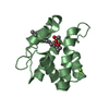
| ||||||||
| Unit cell |
|
-Components
| #1: Protein | / mosquito odorant binding protein Mass: 14396.547 Da / Num. of mol.: 2 Source method: isolated from a genetically manipulated source Source: (gene. exp.) Culex quinquefasciatus (southern house mosquito)Gene: CpipJ_CPIJ007604 / Production host:  Escherichia coli (E. coli) / References: UniProt: Q8T6I2 Escherichia coli (E. coli) / References: UniProt: Q8T6I2#2: Chemical |   Mass: 312.444 Da / Num. of mol.: 2 / Source method: obtained synthetically / Formula: C18H32O4 Mass: 312.444 Da / Num. of mol.: 2 / Source method: obtained synthetically / Formula: C18H32O4#3: Chemical | ChemComp-MG /   Mass: 24.305 Da / Num. of mol.: 4 / Source method: obtained synthetically / Formula: Mg Mass: 24.305 Da / Num. of mol.: 4 / Source method: obtained synthetically / Formula: Mg#4: Water | ChemComp-HOH / | Water Mass: 18.015 Da / Num. of mol.: 419 / Source method: isolated from a natural source / Formula: H2O Mass: 18.015 Da / Num. of mol.: 419 / Source method: isolated from a natural source / Formula: H2O |
|---|
-Experimental details
-Experiment
| Experiment | Method: X-RAY DIFFRACTION / Number of used crystals: 1 |
|---|
- Sample preparation
| Crystal | Density Matthews: 2.54 Å3/Da / Density % sol: 51.51 % |
|---|---|
| Crystal grow | Temperature: 298.15 K / Method: vapor diffusion, hanging drop / pH: 8.2 Details: 20% (wt/vol) PEG 4,000, 100 mM HEPES, 200 mM MgCl2, pH 8.2, VAPOR DIFFUSION, HANGING DROP, temperature 298.15K |
-Data collection
| Diffraction | Mean temperature: 100 K | ||||||||||||||||||||||
|---|---|---|---|---|---|---|---|---|---|---|---|---|---|---|---|---|---|---|---|---|---|---|---|
| Diffraction source | Source: SYNCHROTRON / Site: APS  / Beamline: 24-ID-E / Wavelength: 0.97918 Å / Beamline: 24-ID-E / Wavelength: 0.97918 Å | ||||||||||||||||||||||
| Detector | Type: ADSC QUANTUM 315 / Detector: CCD / Date: Feb 14, 2008 | ||||||||||||||||||||||
| Radiation | Protocol: SINGLE WAVELENGTH / Monochromatic (M) / Laue (L): M / Scattering type: x-ray | ||||||||||||||||||||||
| Radiation wavelength | Wavelength: 0.97918 Å / Relative weight: 1 | ||||||||||||||||||||||
| Reflection | Resolution: 1.3→50 Å / Num. all: 68116 / Num. obs: 68116 / % possible obs: 96.9 % / Observed criterion σ(F): 0 / Observed criterion σ(I): 0 | ||||||||||||||||||||||
| Reflection shell |
|
- Processing
| Software |
| ||||||||||||||||||||||||||||||||||||||||||||||||||||||||||||||||||||||
|---|---|---|---|---|---|---|---|---|---|---|---|---|---|---|---|---|---|---|---|---|---|---|---|---|---|---|---|---|---|---|---|---|---|---|---|---|---|---|---|---|---|---|---|---|---|---|---|---|---|---|---|---|---|---|---|---|---|---|---|---|---|---|---|---|---|---|---|---|---|---|---|
| Refinement | Method to determine structure: MOLECULAR REPLACEMENT / Resolution: 1.3→26.83 Å / Cor.coef. Fo:Fc: 0.972 / Cor.coef. Fo:Fc free: 0.955 / SU B: 1.16 / SU ML: 0.023 / Cross valid method: THROUGHOUT / σ(F): 0 / ESU R Free: 0.047 / Stereochemistry target values: MAXIMUM LIKELIHOOD / Details: HYDROGENS HAVE BEEN ADDED IN THE RIDING POSITIONS
| ||||||||||||||||||||||||||||||||||||||||||||||||||||||||||||||||||||||
| Solvent computation | Ion probe radii: 0.8 Å / Shrinkage radii: 0.8 Å / VDW probe radii: 1.4 Å / Solvent model: MASK | ||||||||||||||||||||||||||||||||||||||||||||||||||||||||||||||||||||||
| Displacement parameters | Biso mean: 16.795 Å2
| ||||||||||||||||||||||||||||||||||||||||||||||||||||||||||||||||||||||
| Refinement step | Cycle: LAST / Resolution: 1.3→26.83 Å
| ||||||||||||||||||||||||||||||||||||||||||||||||||||||||||||||||||||||
| Refine LS restraints |
| ||||||||||||||||||||||||||||||||||||||||||||||||||||||||||||||||||||||
| LS refinement shell | Resolution: 1.3→1.334 Å / Total num. of bins used: 20
|
