 Movie
Movie Controller


+ Open data

- Basic information
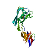
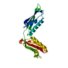
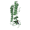
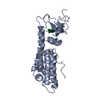
| Entry | Database: PDB / ID: 3og5 | ||||||
|---|---|---|---|---|---|---|---|
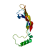
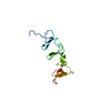
| Title | Crystal Structure of BamA POTRA45 tandem | ||||||
 Components Components | Outer membrane protein assembly complex, YaeT protein | ||||||
 Keywords Keywords |  PROTEIN BINDING / POTRA fold / Insertion of outer membrane proteins PROTEIN BINDING / POTRA fold / Insertion of outer membrane proteins | ||||||
| Function / homology |  Function and homology information Function and homology informationBam protein complex / Gram-negative-bacterium-type cell outer membrane assembly / protein insertion into membrane / cell outer membrane / cell adhesion / membraneSimilarity search - Function | ||||||
| Biological species |  Escherichia coli (E. coli) Escherichia coli (E. coli) | ||||||
| Method | X-RAY DIFFRACTION / SYNCHROTRON / SAD / Resolution: 2.69 Å | ||||||
 Authors Authors | Gatzeva-Topalova, P.Z. / Warner, L.R. / Pardi, A. / Sousa, M.C. | ||||||
 Citation Citation | Journal: Structure / Year: 2010 Title: Structure and Flexibility of the Complete Periplasmic Domain of BamA: The Protein Insertion Machine of the Outer Membrane Authors: Gatzeva-Topalova, P.Z. / Warner, L.R. / Pardi, A. / Sousa, M.C. | ||||||
| History |
|
- Structure visualization
| Structure viewer | Molecule: MolmilJmol/JSmol |
|---|
- Downloads & links
-Download
| PDBx/mmCIF format | 3og5.cif.gz | 125.5 KB | Display | PDBx/mmCIF format |
|---|---|---|---|---|
| PDB format | pdb3og5.ent.gz | 103.9 KB | Display | PDB format |
| PDBx/mmJSON format | 3og5.json.gz | Tree view | PDBx/mmJSON format | |
| Others |  Other downloads Other downloads |
-Validation report
| Arichive directory | https://data.pdbj.org/pub/pdb/validation_reports/og/3og5ftp://data.pdbj.org/pub/pdb/validation_reports/og/3og5 | HTTPS FTP |
|---|
-Related structure data
| Similar structure data |
|---|


-Links
 PDBj
PDBj- Assembly
| Deposited unit | 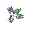
| ||||||||
|---|---|---|---|---|---|---|---|---|---|
| 1 | 
| ||||||||
| 2 | 
| ||||||||
| Unit cell |
|
-Components
| #1: Protein | Mass: 18936.482 Da / Num. of mol.: 2 / Fragment: POTRA45 DOMAIN (unp residues 264-424) Source method: isolated from a genetically manipulated source Source: (gene. exp.) Escherichia coli (E. coli) / Strain: K12 / Gene: yaeT / Plasmid: pMS487 / Production host: Escherichia coli (E. coli) / Strain (production host): Rosetta DE3 / References: UniProt: D5CVA9, UniProt: P0A940*PLUS#2: Water | ChemComp-HOH / | Water Mass: 18.015 Da / Num. of mol.: 50 / Source method: isolated from a natural source / Formula: H2O Mass: 18.015 Da / Num. of mol.: 50 / Source method: isolated from a natural source / Formula: H2O |
|---|
-Experimental details
-Experiment
| Experiment | Method: X-RAY DIFFRACTION / Number of used crystals: 1 |
|---|
- Sample preparation
| Crystal | Density Matthews: 3.75 Å3/Da / Density % sol: 67.16 % |
|---|---|
| Crystal grow | Temperature: 289 K / Method: vapor diffusion, hanging drop / pH: 7.5 Details: 0.1M HEPES pH 7.5, 1.68M ammonium sulfate, 3% PEG 400, 0.25M NaCl, VAPOR DIFFUSION, HANGING DROP, temperature 289K |
-Data collection
| Diffraction | Mean temperature: 100 K | ||||||||||||||||||||||||||||||||||||||||||||||||||||||||||||||||||
|---|---|---|---|---|---|---|---|---|---|---|---|---|---|---|---|---|---|---|---|---|---|---|---|---|---|---|---|---|---|---|---|---|---|---|---|---|---|---|---|---|---|---|---|---|---|---|---|---|---|---|---|---|---|---|---|---|---|---|---|---|---|---|---|---|---|---|---|
| Diffraction source | Source: SYNCHROTRON / Site: ALS  / Beamline: 8.2.2 / Wavelength: 0.9791 Å / Beamline: 8.2.2 / Wavelength: 0.9791 Å | ||||||||||||||||||||||||||||||||||||||||||||||||||||||||||||||||||
| Detector | Type: ADSC QUANTUM 315 / Detector: CCD / Date: Oct 23, 2008 | ||||||||||||||||||||||||||||||||||||||||||||||||||||||||||||||||||
| Radiation | Protocol: SINGLE WAVELENGTH / Monochromatic (M) / Laue (L): M / Scattering type: x-ray | ||||||||||||||||||||||||||||||||||||||||||||||||||||||||||||||||||
| Radiation wavelength | Wavelength: 0.9791 Å / Relative weight: 1 | ||||||||||||||||||||||||||||||||||||||||||||||||||||||||||||||||||
| Reflection | Resolution: 2.69→40 Å / Num. obs: 29399 / % possible obs: 96.8 % / Redundancy: 1.5 % / Rmerge(I) obs: 0.084 / Χ2: 1.34 / Net I/σ(I): 8.4 | ||||||||||||||||||||||||||||||||||||||||||||||||||||||||||||||||||
| Reflection shell |
|
- Processing
| Software |
| |||||||||||||||||||||||||||||||||||||||||||||||||||||||||||||||||||||||||||||
|---|---|---|---|---|---|---|---|---|---|---|---|---|---|---|---|---|---|---|---|---|---|---|---|---|---|---|---|---|---|---|---|---|---|---|---|---|---|---|---|---|---|---|---|---|---|---|---|---|---|---|---|---|---|---|---|---|---|---|---|---|---|---|---|---|---|---|---|---|---|---|---|---|---|---|---|---|---|---|
| Refinement | Method to determine structure: SAD / Resolution: 2.69→38.786 Å / Occupancy max: 1 / Occupancy min: 1 / FOM work R set: 0.8206 / SU ML: 0.4 / σ(F): 0.08 / Phase error: 25.21 / Stereochemistry target values: ML
| |||||||||||||||||||||||||||||||||||||||||||||||||||||||||||||||||||||||||||||
| Solvent computation | Shrinkage radii: 0.9 Å / VDW probe radii: 1.11 Å / Solvent model: FLAT BULK SOLVENT MODEL / Bsol: 27.374 Å2 / ksol: 0.333 e/Å3 | |||||||||||||||||||||||||||||||||||||||||||||||||||||||||||||||||||||||||||||
| Displacement parameters | Biso max: 218.24 Å2 / Biso mean: 42.8419 Å2 / Biso min: 2.62 Å2
| |||||||||||||||||||||||||||||||||||||||||||||||||||||||||||||||||||||||||||||
| Refinement step | Cycle: LAST / Resolution: 2.69→38.786 Å
| |||||||||||||||||||||||||||||||||||||||||||||||||||||||||||||||||||||||||||||
| Refine LS restraints |
| |||||||||||||||||||||||||||||||||||||||||||||||||||||||||||||||||||||||||||||
| LS refinement shell | Refine-ID: X-RAY DIFFRACTION / Total num. of bins used: 10
|
