 Movie
Movie Controller

[English] 日本語
 Yorodumi
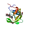
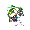
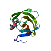
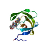
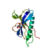
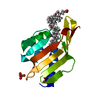
- PDB-3obl: Crystal structure of the potent anti-HIV cyanobacterial lectin fr... -
+ Open data

- Basic information
| Entry | Database: PDB / ID: 3obl | ||||||
|---|---|---|---|---|---|---|---|
| Title | Crystal structure of the potent anti-HIV cyanobacterial lectin from Oscillatoria Agardhii | ||||||
 Components Components | Lectin | ||||||
 Keywords Keywords | SUGAR BINDING PROTEIN / novel beta barrel fold / anti-HIV lectin / High mannose glycans | ||||||
| Function / homology | Lipocalin - #450 / OAA-family lectin sugar binding domain / OAA-family lectin sugar binding domain / Lipocalin / carbohydrate binding / Beta Barrel / Mainly Beta / Lectin Function and homology information Function and homology information | ||||||
| Biological species |  Planktothrix agardhii (bacteria) Planktothrix agardhii (bacteria) | ||||||
| Method | X-RAY DIFFRACTION / SYNCHROTRON / MAD / Resolution: 1.2 Å | ||||||
 Authors Authors | Koharudin, L.M.I. / Furey, W. / Gronenborn, A.M. | ||||||
 Citation Citation | Journal: J.Biol.Chem. / Year: 2011 Title: Novel fold and carbohydrate specificity of the potent anti-HIV cyanobacterial lectin from Oscillatoria agardhii. Authors: Koharudin, L.M. / Furey, W. / Gronenborn, A.M. #1: Journal: J.Biol.Chem. / Year: 2007 Title: Primary structure and carbohydrate binding specificity of a potent anti-HIV lectin isolated from the filamentous cyanobacterium Oscillatoria agardhii Authors: Sato, Y. / Okuyama, S. / Hori, K. #2: Journal: Fisheries Sci. / Year: 2009Title: Cloning, expression, and characterization of a novel anti-HIV lectin from the cultured cyanobacterium, Oscillatoria agardhii Authors: Sato, T. / Hori, K. | ||||||
| History |
|
- Structure visualization
| Structure viewer | Molecule: MolmilJmol/JSmol |
|---|
- Downloads & links
-Download
| PDBx/mmCIF format | 3obl.cif.gz | 133.6 KB | Display | PDBx/mmCIF format |
|---|---|---|---|---|
| PDB format | pdb3obl.ent.gz | 105.7 KB | Display | PDB format |
| PDBx/mmJSON format | 3obl.json.gz | Tree view | PDBx/mmJSON format | |
| Others |  Other downloads Other downloads |
-Validation report
| Arichive directory | https://data.pdbj.org/pub/pdb/validation_reports/ob/3oblftp://data.pdbj.org/pub/pdb/validation_reports/ob/3obl | HTTPS FTP |
|---|
-Related structure data
| Similar structure data |
|---|


-Links
 PDBj
PDBj- Assembly
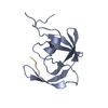
| Deposited unit | 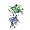
| ||||||||
|---|---|---|---|---|---|---|---|---|---|
| 1 | 
| ||||||||
| 2 | 
| ||||||||
| Unit cell |
|
-Components
| #1: Protein | Mass: 13930.758 Da / Num. of mol.: 2 Source method: isolated from a genetically manipulated source Details: Expressed as native protein / Source: (gene. exp.) Planktothrix agardhii (bacteria) / Gene: OAA / Plasmid: pET26B(+) / Production host: Escherichia coli (E. coli) / Strain (production host): Rosetta 2 (DE3) / References: UniProt: C0STD7#2: Chemical | ChemComp-CXS / CAPS (buffer)  Mass: 221.317 Da / Num. of mol.: 4 / Source method: obtained synthetically / Formula: C9H19NO3S / Comment: pH buffer*YM Mass: 221.317 Da / Num. of mol.: 4 / Source method: obtained synthetically / Formula: C9H19NO3S / Comment: pH buffer*YM#3: Water | ChemComp-HOH / | Water Mass: 18.015 Da / Num. of mol.: 344 / Source method: isolated from a natural source / Formula: H2O Mass: 18.015 Da / Num. of mol.: 344 / Source method: isolated from a natural source / Formula: H2O |
|---|
-Experimental details
-Experiment
| Experiment | Method: X-RAY DIFFRACTION / Number of used crystals: 1 |
|---|
- Sample preparation
| Crystal | Density Matthews: 2.71 Å3/Da / Density % sol: 54.54 % |
|---|---|
| Crystal grow | Temperature: 298 K / Method: vapor diffusion, sitting drop / pH: 8 Details: 1.2 M NaH2PO4/0.8 M K2HPO4, 0.2 M Li2SO4, 0.1 M CAPS (pH 10.5), VAPOR DIFFUSION, SITTING DROP, temperature 298K |
-Data collection
| Diffraction | Mean temperature: 277 K | ||||||||||||
|---|---|---|---|---|---|---|---|---|---|---|---|---|---|
| Diffraction source | Source: SYNCHROTRON / Site: APS  / Beamline: 22-BM / Wavelength: 0.9795, 0.9793, 0.9718 / Beamline: 22-BM / Wavelength: 0.9795, 0.9793, 0.9718 | ||||||||||||
| Detector | Type: MAR scanner 300 mm plate / Detector: IMAGE PLATE / Date: Feb 25, 2010 / Details: MIRRORS | ||||||||||||
| Radiation | Monochromator: ROSENBAUM-ROCK MONOCHROMATOR HIGH-RESOLUTION DOUBLE-CRYSTAL SI(220) SAGITTAL FOCUSING, ROSENBAUM- ROCK VERTICAL FOCUSING MIRROR Protocol: MAD / Monochromatic (M) / Laue (L): M / Scattering type: x-ray | ||||||||||||
| Radiation wavelength |
| ||||||||||||
| Reflection | Resolution: 1.1→22.64 Å / Num. obs: 86175 / % possible obs: 92 % / Observed criterion σ(F): 1 / Observed criterion σ(I): 3 / Redundancy: 3.81 % / Rmerge(I) obs: 0.079 / Net I/σ(I): 7.5 | ||||||||||||
| Reflection shell | Resolution: 1.1→1.14 Å / Redundancy: 3.73 % / Rmerge(I) obs: 0.434 / Mean I/σ(I) obs: 2.1 / Num. unique all: 9892 / % possible all: 83.1 |
- Processing
| Software |
| ||||||||||||||||||||||||||||||||||||||||||||||||||||||||||||||||||||||||||||||||||||||||||||||||||||
|---|---|---|---|---|---|---|---|---|---|---|---|---|---|---|---|---|---|---|---|---|---|---|---|---|---|---|---|---|---|---|---|---|---|---|---|---|---|---|---|---|---|---|---|---|---|---|---|---|---|---|---|---|---|---|---|---|---|---|---|---|---|---|---|---|---|---|---|---|---|---|---|---|---|---|---|---|---|---|---|---|---|---|---|---|---|---|---|---|---|---|---|---|---|---|---|---|---|---|---|---|---|
| Refinement | Method to determine structure: MAD / Resolution: 1.2→22.64 Å / Cor.coef. Fo:Fc: 0.976 / Cor.coef. Fo:Fc free: 0.967 / SU B: 1.692 / SU ML: 0.033 / Isotropic thermal model: Anisotropic / Cross valid method: THROUGHOUT / σ(F): 1 / ESU R Free: 0.039 / Stereochemistry target values: MAXIMUM LIKELIHOOD Details: HYDROGENS HAVE BEEN ADDED IN THE RIDING POSITIONS. THE DATA HAVE BEEN COLLECTED UP 1.10 A RESOLUTION. HOWEVER, REFINEMENT CONVERGED TO BETTER R VALUES BY CUTTING THE RESOLUTION TO 1.20 A.
| ||||||||||||||||||||||||||||||||||||||||||||||||||||||||||||||||||||||||||||||||||||||||||||||||||||
| Solvent computation | Ion probe radii: 0.8 Å / Shrinkage radii: 0.8 Å / VDW probe radii: 1.4 Å / Solvent model: BABINET MODEL WITH MASK | ||||||||||||||||||||||||||||||||||||||||||||||||||||||||||||||||||||||||||||||||||||||||||||||||||||
| Displacement parameters | Biso mean: 13.854 Å2
| ||||||||||||||||||||||||||||||||||||||||||||||||||||||||||||||||||||||||||||||||||||||||||||||||||||
| Refinement step | Cycle: LAST / Resolution: 1.2→22.64 Å
| ||||||||||||||||||||||||||||||||||||||||||||||||||||||||||||||||||||||||||||||||||||||||||||||||||||
| Refine LS restraints |
| ||||||||||||||||||||||||||||||||||||||||||||||||||||||||||||||||||||||||||||||||||||||||||||||||||||
| LS refinement shell | Resolution: 1.2→1.231 Å / Total num. of bins used: 20
|
