[English] 日本語
Yorodumi
- PDB-3o8s: Crystal structure of an ADP-ribose pyrophosphatase (SSU98_1448) f... -

+
Open data


ID or keywords:

Loading...

-
Basic information

Entry
Database: PDB / ID: 3o8s
TitleCrystal structure of an ADP-ribose pyrophosphatase (SSU98_1448) from STREPTOCOCCUS SUIS 89-1591 at 2.27 A resolution
ComponentsADP-ribose pyrophosphatase
KeywordsHYDROLASE / STRUCTURAL GENOMICS / JOINT CENTER FOR STRUCTURAL GENOMICS / JCSG / PROTEIN STRUCTURE INITIATIVE / PSI-BIOLOGY / NUDIX PROTEIN
Function / homologyNucleoside Triphosphate Pyrophosphohydrolase / Nucleoside Triphosphate Pyrophosphohydrolase / Alpha-Beta Complex / Alpha Beta / ACETATE ION / :
Function and homology information
Biological speciesStreptococcus suis (bacteria)
MethodX-RAY DIFFRACTION / SYNCHROTRON / MOLECULAR REPLACEMENT / molecular replacement / Resolution: 2.27 Å
AuthorsJoint Center for Structural Genomics (JCSG)
CitationJournal: To be published
Title: Crystal structure of an ADP-ribose pyrophosphatase (SSU98_1448) from STREPTOCOCCUS SUIS 89-1591 at 2.27 A resolution
Authors: Joint Center for Structural Genomics (JCSG)
History
DepositionAug 3, 2010Deposition site: RCSB / Processing site: RCSB
Revision 1.0Aug 25, 2010Provider: repository / Type: Initial release
Revision 1.1Jul 13, 2011Group: Version format compliance
Revision 1.2Jul 20, 2011Group: Structure summary
Revision 1.3Feb 1, 2023Group: Database references / Derived calculations / Category: database_2 / struct_ref_seq_dif / struct_site
Item: _database_2.pdbx_DOI / _database_2.pdbx_database_accession ..._database_2.pdbx_DOI / _database_2.pdbx_database_accession / _struct_ref_seq_dif.details / _struct_site.pdbx_auth_asym_id / _struct_site.pdbx_auth_comp_id / _struct_site.pdbx_auth_seq_id

-
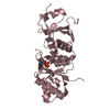
Structure visualization

Structure viewerMolecule:
MolmilJmol/JSmol

Downloads & links

-
Assembly

Deposited unit
A: ADP-ribose pyrophosphatase
hetero molecules


Theoretical massNumber of molelcules
Total (without water)23,7357
Polymers23,3661
Non-polymers3696
Water63135
1
A: ADP-ribose pyrophosphatase
hetero molecules

A: ADP-ribose pyrophosphatase
hetero molecules


Theoretical massNumber of molelcules
Total (without water)47,47114
Polymers46,7322
Non-polymers73912
Water362
TypeNameSymmetry operationNumber
identity operation1_555x,y,z1
crystal symmetry operation8_665-y+1,-x+1,-z+3/41
Buried area4080 Å2
ΔGint10 kcal/mol
Surface area12790 Å2
MethodPISA
Unit cell
Length a, b, c (Å)64.780, 64.780, 108.290
Angle α, β, γ (deg.)90.000, 90.000, 90.000
Int Tables number95
Space group name H-MP4322

-
Components

#1: Protein ADP-ribose pyrophosphatase / NUDIX hydrolase


Mass: 23365.881 Da / Num. of mol.: 1
Source method: isolated from a genetically manipulated source
Source: (gene. exp.) Streptococcus suis (bacteria) / Strain: 89/1591 / Gene: SsuiDRAFT_3457 / Plasmid: SpeedET / Production host: Escherichia Coli (E. coli) / Strain (production host): HK100 / References: UniProt: B9WTJ0
#2: Chemical
ChemComp-EDO / 1,2-ETHANEDIOL / ETHYLENE GLYCOL / Ethylene glycol


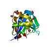
Mass: 62.068 Da / Num. of mol.: 5 / Source method: obtained synthetically / Formula: C2H6O2
#3: Chemical ChemComp-ACT / ACETATE ION / Acetate


Mass: 59.044 Da / Num. of mol.: 1 / Source method: obtained synthetically / Formula: C2H3O2
#4: Water ChemComp-HOH / water / Water


Mass: 18.015 Da / Num. of mol.: 35 / Source method: isolated from a natural source / Formula: H2O
Sequence detailsTHE CONSTRUCT WAS EXPRESSED WITH A PURIFICATION TAG MGSDKIHHHHHHENLYFQG. THE TAG WAS REMOVED WITH ...THE CONSTRUCT WAS EXPRESSED WITH A PURIFICATION TAG MGSDKIHHHHHHENLYFQG. THE TAG WAS REMOVED WITH TEV PROTEASE LEAVING ONLY A GLYCINE (0) FOLLOWED BY THE TARGET SEQUENCE.

-
Experimental details

-
Experiment

ExperimentMethod: X-RAY DIFFRACTION / Number of used crystals: 1

-
Sample preparation

CrystalDensity Matthews: 2.43 Å3/Da / Density % sol: 49.4 %
Description: DATA WERE SCALED USING XSCALE WITH FRIEDEL PAIRS KEPT AS SEPARATE WHEN COMPUTING R-MERGE, COMPLETENESS AND
Crystal growTemperature: 293 K / Method: vapor diffusion, sitting drop / pH: 4.5
Details: 50.0% ethylene glycol, 5.0% polyethylene glycol 1000, 0.1M sodium acetate pH 4.5, NANODROP, VAPOR DIFFUSION, SITTING DROP, temperature 293K

-
Data collection

DiffractionMean temperature: 100 K
Diffraction sourceSource: SYNCHROTRON / Site: SSRL / Beamline: BL11-1 / Wavelength: 0.97885
DetectorType: MARMOSAIC 325 mm CCD / Detector: CCD / Date: Apr 8, 2007 / Details: Flat mirror (vertical focusing)
RadiationMonochromator: Single crystal Si(111) bent monochromator (horizontal focusing)
Protocol: SINGLE WAVELENGTH / Monochromatic (M) / Laue (L): M / Scattering type: x-ray
Radiation wavelengthWavelength: 0.97885 Å / Relative weight: 1
ReflectionResolution: 2.27→28.97 Å / Num. obs: 10944 / % possible obs: 97.7 % / Observed criterion σ(I): -3 / Biso Wilson estimate: 52.96 Å2 / Rmerge(I) obs: 0.05 / Net I/σ(I): 17.09
Reflection shell

Diffraction-ID: 1

Resolution (Å)Highest resolution (Å)Rmerge(I) obsMean I/σ(I) obsNum. measured obsNum. unique obs% possible all
2.27-2.350.7612.37656198298.9
2.35-2.440.572.97533194599.1
2.44-2.560.448383215598.2
2.56-2.690.2865.77620195298.2
2.69-2.860.18587840201098.5
2.86-3.080.1112.67838199998.1
3.08-3.390.06419.97872200297.8
3.39-3.870.03929.67621193197.4
3.87-4.870.02641.67902199896.9
4.870.02445.77765196194

-
Phasing

PhasingMethod: molecular replacement

-
Processing

Software
NameVersionClassificationNB
BUSTER-TNTBUSTER 2.8.0refinement
XSCALEdata processing
PDB_EXTRACT3.1data extraction
XDSdata reduction
XSCALEdata scaling
Rosettaphasing
PHASERphasing
BUSTER2.8.0refinement
RefinementMethod to determine structure: MOLECULAR REPLACEMENT / Resolution: 2.27→28.97 Å / Cor.coef. Fo:Fc: 0.9475 / Cor.coef. Fo:Fc free: 0.9375 / Occupancy max: 1 / Occupancy min: 0.5 / Cross valid method: THROUGHOUT / σ(F): 0
Details: 1. 1,2-ETHANEDIOL (EDO) AND ACETATE (ACT) FROM THE CRYSTALLIZATION CONDITION ARE MODELED INTO THE STRUCTURE. 2. ATOM RECORD CONTAINS SUM OF TLS AND RESIDUAL B FACTORS. ANISOU RECORD CONTAINS ...Details: 1. 1,2-ETHANEDIOL (EDO) AND ACETATE (ACT) FROM THE CRYSTALLIZATION CONDITION ARE MODELED INTO THE STRUCTURE. 2. ATOM RECORD CONTAINS SUM OF TLS AND RESIDUAL B FACTORS. ANISOU RECORD CONTAINS SUM OF TLS AND RESIDUAL U FACTORS. 3. ELECTRON DENSITY FOR RESIDUES 1-66 IN THE N-TERMINAL REGION OF THE PROTEIN IS DISORDERED, AND THIS REGION COULD NOT BE MODELED.
RfactorNum. reflection% reflectionSelection details
Rfree0.2179 959 8.78 %RANDOM
Rwork0.1906 ---
obs0.1931 10920 --
Displacement parametersBiso max: 122.35 Å2 / Biso mean: 61.3391 Å2 / Biso min: 32.57 Å2
Baniso -1Baniso -2Baniso -3
1--3.4675 Å20 Å20 Å2
2---3.4675 Å20 Å2
3---6.9349 Å2
Refinement stepCycle: LAST / Resolution: 2.27→28.97 Å
ProteinNucleic acidLigandSolventTotal
Num. atoms1046 0 24 35 1105
Refine LS restraints
Refine-IDTypeNumberRestraint functionWeightDev ideal
X-RAY DIFFRACTIONt_dihedral_angle_d499SINUSOIDAL2
X-RAY DIFFRACTIONt_trig_c_planes37HARMONIC2
X-RAY DIFFRACTIONt_gen_planes149HARMONIC5
X-RAY DIFFRACTIONt_it1083HARMONIC20
X-RAY DIFFRACTIONt_nbd
X-RAY DIFFRACTIONt_improper_torsion
X-RAY DIFFRACTIONt_pseud_angle
X-RAY DIFFRACTIONt_chiral_improper_torsion136SEMIHARMONIC5
X-RAY DIFFRACTIONt_sum_occupancies
X-RAY DIFFRACTIONt_utility_distance
X-RAY DIFFRACTIONt_utility_angle
X-RAY DIFFRACTIONt_utility_torsion
X-RAY DIFFRACTIONt_ideal_dist_contact1203SEMIHARMONIC4
X-RAY DIFFRACTIONt_bond_d1083HARMONIC20.01
X-RAY DIFFRACTIONt_angle_deg1460HARMONIC21.07
X-RAY DIFFRACTIONt_omega_torsion3.69
X-RAY DIFFRACTIONt_other_torsion2.87
LS refinement shellResolution: 2.27→2.49 Å / Total num. of bins used: 6
RfactorNum. reflection% reflection
Rfree0.2144 192 7.42 %
Rwork0.192 2394 -
all0.1937 2586 -
Refinement TLS params.Method: refined / Origin x: 16.6105 Å / Origin y: 36.7788 Å / Origin z: 51.912 Å
111213212223313233
T-0.2379 Å20.0425 Å20.0089 Å2-0.1112 Å20.0088 Å2---0.1745 Å2
L3.1357 °20.0389 °2-0.5016 °2-1.0587 °2-0.4393 °2--6.8452 °2
S-0.0629 Å °-0.4622 Å °0.047 Å °-0.0718 Å °0.0957 Å °0.2468 Å °-0.1676 Å °-1.08 Å °-0.0328 Å °
Refinement TLS groupSelection details: { A|* }

+
About Yorodumi

-
News

-
Feb 9, 2022. New format data for meta-information of EMDB entries

New format data for meta-information of EMDB entries

  • Version 3 of the EMDB header file is now the official format.
  • The previous official version 1.9 will be removed from the archive.

Related info.:EMDB header

External links:wwPDB to switch to version 3 of the EMDB data model

-
Aug 12, 2020. Covid-19 info

Covid-19 info

URL: https://pdbj.org/emnavi/covid19.php

New page: Covid-19 featured information page in EM Navigator.

Related info.:Covid-19 info / Mar 5, 2020. Novel coronavirus structure data

+
Mar 5, 2020. Novel coronavirus structure data

Novel coronavirus structure data

Related info.:Yorodumi Speices / Aug 12, 2020. Covid-19 info

External links:COVID-19 featured content - PDBj / Molecule of the Month (242):Coronavirus Proteases

+
Jan 31, 2019. EMDB accession codes are about to change! (news from PDBe EMDB page)

EMDB accession codes are about to change! (news from PDBe EMDB page)

  • The allocation of 4 digits for EMDB accession codes will soon come to an end. Whilst these codes will remain in use, new EMDB accession codes will include an additional digit and will expand incrementally as the available range of codes is exhausted. The current 4-digit format prefixed with “EMD-” (i.e. EMD-XXXX) will advance to a 5-digit format (i.e. EMD-XXXXX), and so on. It is currently estimated that the 4-digit codes will be depleted around Spring 2019, at which point the 5-digit format will come into force.
  • The EM Navigator/Yorodumi systems omit the EMD- prefix.

Related info.:Q: What is EMD? / ID/Accession-code notation in Yorodumi/EM Navigator

External links:EMDB Accession Codes are Changing Soon! / Contact to PDBj

+
Jul 12, 2017. Major update of PDB

Major update of PDB

  • wwPDB released updated PDB data conforming to the new PDBx/mmCIF dictionary.
  • This is a major update changing the version number from 4 to 5, and with Remediation, in which all the entries are updated.
  • In this update, many items about electron microscopy experimental information are reorganized (e.g. em_software).
  • Now, EM Navigator and Yorodumi are based on the updated data.

External links:wwPDB Remediation / Enriched Model Files Conforming to OneDep Data Standards Now Available in the PDB FTP Archive

-
Yorodumi

Thousand views of thousand structures

  • Yorodumi is a browser for structure data from EMDB, PDB, SASBDB, etc.
  • This page is also the successor to EM Navigator detail page, and also detail information page/front-end page for Omokage search.
  • The word "yorodu" (or yorozu) is an old Japanese word meaning "ten thousand". "mi" (miru) is to see.

Related info.:EMDB / PDB / SASBDB / Comparison of 3 databanks / Yorodumi Search / Aug 31, 2016. New EM Navigator & Yorodumi / Yorodumi Papers / Jmol/JSmol / Function and homology information / Changes in new EM Navigator and Yorodumi

Read more