 Movie
Movie Controller


+ Open data

- Basic information
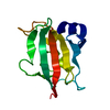
| Entry | Database: PDB / ID: 3o5o | ||||||
|---|---|---|---|---|---|---|---|
| Title | Fk1 domain mutant A19T of FKBP51, crystal form III | ||||||
 Components Components | Peptidyl-prolyl cis-trans isomerase FKBP5 | ||||||
 Keywords Keywords |  ISOMERASE / Fk-506 binding domain / Hsp90 cochaperone / immunophiline / peptidyl-prolyl isomerase ISOMERASE / Fk-506 binding domain / Hsp90 cochaperone / immunophiline / peptidyl-prolyl isomerase | ||||||
| Function / homology |  Function and homology informationFK506 binding / chaperone-mediated protein folding / MECP2 regulates neuronal receptors and channels / heat shock protein binding / HSP90 chaperone cycle for steroid hormone receptors (SHR) in the presence of ligand / ESR-mediated signaling / peptidylprolyl isomerase / peptidyl-prolyl cis-trans isomerase activity / response to bacterium / protein folding ...FK506 binding / chaperone-mediated protein folding / MECP2 regulates neuronal receptors and channels / heat shock protein binding / HSP90 chaperone cycle for steroid hormone receptors (SHR) in the presence of ligand / ESR-mediated signaling / peptidylprolyl isomerase / peptidyl-prolyl cis-trans isomerase activity / response to bacterium / protein folding / extracellular exosome / nucleoplasm / membrane / cytosol / cytoplasm Function and homology informationFK506 binding / chaperone-mediated protein folding / MECP2 regulates neuronal receptors and channels / heat shock protein binding / HSP90 chaperone cycle for steroid hormone receptors (SHR) in the presence of ligand / ESR-mediated signaling / peptidylprolyl isomerase / peptidyl-prolyl cis-trans isomerase activity / response to bacterium / protein folding ...FK506 binding / chaperone-mediated protein folding / MECP2 regulates neuronal receptors and channels / heat shock protein binding / HSP90 chaperone cycle for steroid hormone receptors (SHR) in the presence of ligand / ESR-mediated signaling / peptidylprolyl isomerase / peptidyl-prolyl cis-trans isomerase activity / response to bacterium / protein folding / extracellular exosome / nucleoplasm / membrane / cytosol / cytoplasmSimilarity search - Function | ||||||
| Biological species |  Homo sapiens (human) Homo sapiens (human) | ||||||
| Method | X-RAY DIFFRACTION / SYNCHROTRON / MOLECULAR REPLACEMENT / molecular replacement / Resolution: 1.15 Å | ||||||
 Authors Authors | Bracher, A. / Kozany, C. / Thost, A.-K. / Hausch, F. | ||||||
 Citation Citation | Journal: Acta Crystallogr.,Sect.D / Year: 2011 Title: Structural characterization of the PPIase domain of FKBP51, a cochaperone of human Hsp90. Authors: Bracher, A. / Kozany, C. / Thost, A.K. / Hausch, F. | ||||||
| History |
|
- Structure visualization
| Structure viewer | Molecule: MolmilJmol/JSmol |
|---|
- Downloads & links
-Download
| PDBx/mmCIF format | 3o5o.cif.gz | 76.1 KB | Display | PDBx/mmCIF format |
|---|---|---|---|---|
| PDB format | pdb3o5o.ent.gz | 54.7 KB | Display | PDB format |
| PDBx/mmJSON format | 3o5o.json.gz | Tree view | PDBx/mmJSON format | |
| Others |  Other downloads Other downloads |
-Validation report
| Arichive directory | https://data.pdbj.org/pub/pdb/validation_reports/o5/3o5oftp://data.pdbj.org/pub/pdb/validation_reports/o5/3o5o | HTTPS FTP |
|---|
-Related structure data
| Related structure data |  3o5dC  3o5eC  3o5fC  3o5gC  3o5iC  3o5jC  3o5kC  3o5lC  3o5mC  3o5pSC  3o5qC  3o5rC C: citing same article ( S: Starting model for refinement |
|---|---|
| Similar structure data |


-Links
 PDBj
PDBj


- Assembly
| Deposited unit | 
| ||||||||
|---|---|---|---|---|---|---|---|---|---|
| 1 |
| ||||||||
| Unit cell |
|
-Components
| #1: Protein | Mass: 14026.077 Da / Num. of mol.: 1 / Mutation: A19T Source method: isolated from a genetically manipulated source Source: (gene. exp.) Homo sapiens (human) / Gene: AIG6, FKBP5, FKBP51 / Plasmid: pProEx-HtB / Production host:  Escherichia coli (E. coli) / Strain (production host): BL21(DE3) codon+ RIL / References: UniProt: Q13451, peptidylprolyl isomerase Escherichia coli (E. coli) / Strain (production host): BL21(DE3) codon+ RIL / References: UniProt: Q13451, peptidylprolyl isomerase |
|---|---|
| #2: Chemical | ChemComp-SCN / Thiocyanate  Mass: 58.082 Da / Num. of mol.: 1 / Source method: obtained synthetically / Formula: CNS Mass: 58.082 Da / Num. of mol.: 1 / Source method: obtained synthetically / Formula: CNS |
| #3: Water | ChemComp-HOH / Water Mass: 18.015 Da / Num. of mol.: 215 / Source method: isolated from a natural source / Formula: H2O Mass: 18.015 Da / Num. of mol.: 215 / Source method: isolated from a natural source / Formula: H2O |
| Sequence details | AUTHORS STATE THAT A19T IS A MUTATION, BUT NOT A NATURALLY OCCURRING ALLELE. |
-Experimental details
-Experiment
| Experiment | Method: X-RAY DIFFRACTION / Number of used crystals: 1 |
|---|
- Sample preparation
| Crystal | Density Matthews: 1.93 Å3/Da / Density % sol: 36.34 % |
|---|---|
| Crystal grow | Temperature: 293 K / Method: vapor diffusion / pH: 6.5 Details: 33 % PEG3350, 0.05 M KSCN, 0.1 M BisTrisHCl, pH 6.5, vapor diffusion, temperature 293K |
-Data collection
| Diffraction | Mean temperature: 100 K | ||||||||||||||||||||||||||||||||||||||||||||||||||||||||||||||||||||||||||||||||||||||||
|---|---|---|---|---|---|---|---|---|---|---|---|---|---|---|---|---|---|---|---|---|---|---|---|---|---|---|---|---|---|---|---|---|---|---|---|---|---|---|---|---|---|---|---|---|---|---|---|---|---|---|---|---|---|---|---|---|---|---|---|---|---|---|---|---|---|---|---|---|---|---|---|---|---|---|---|---|---|---|---|---|---|---|---|---|---|---|---|---|---|
| Diffraction source | Source: SYNCHROTRON / Site: SLS  / Beamline: X10SA / Wavelength: 0.9794 Å / Beamline: X10SA / Wavelength: 0.9794 Å | ||||||||||||||||||||||||||||||||||||||||||||||||||||||||||||||||||||||||||||||||||||||||
| Detector | Type: MARRESEARCH / Detector: CCD / Date: Dec 18, 2006 | ||||||||||||||||||||||||||||||||||||||||||||||||||||||||||||||||||||||||||||||||||||||||
| Radiation | Protocol: SINGLE WAVELENGTH / Monochromatic (M) / Laue (L): M / Scattering type: x-ray | ||||||||||||||||||||||||||||||||||||||||||||||||||||||||||||||||||||||||||||||||||||||||
| Radiation wavelength | Wavelength: 0.9794 Å / Relative weight: 1 | ||||||||||||||||||||||||||||||||||||||||||||||||||||||||||||||||||||||||||||||||||||||||
| Reflection | Resolution: 1.15→38.296 Å / Num. obs: 38609 / % possible obs: 98.3 % / Redundancy: 4.9 % / Rmerge(I) obs: 0.047 / Rsym value: 0.047 / Net I/σ(I): 17.7 | ||||||||||||||||||||||||||||||||||||||||||||||||||||||||||||||||||||||||||||||||||||||||
| Reflection shell | Diffraction-ID: 1
|
-Phasing
| Phasing | Method: molecular replacement | |||||||||
|---|---|---|---|---|---|---|---|---|---|---|
| Phasing MR | Rfactor: 0.449 / Cor.coef. Fo:Fc: 0.512
|
- Processing
| Software |
| |||||||||||||||||||||||||||||||||
|---|---|---|---|---|---|---|---|---|---|---|---|---|---|---|---|---|---|---|---|---|---|---|---|---|---|---|---|---|---|---|---|---|---|---|
| Refinement | Method to determine structure: MOLECULAR REPLACEMENT Starting model: PDB entry 3O5P Resolution: 1.15→20 Å / Num. parameters: 11299 / Num. restraintsaints: 14104 / Occupancy max: 1 / Occupancy min: 0.32 / Cross valid method: FREE R / σ(F): 0 / Stereochemistry target values: ENGH AND HUBER
| |||||||||||||||||||||||||||||||||
| Solvent computation | Solvent model: MOEWS & KRETSINGER, J.MOL.BIOL.91(1973)201-228 | |||||||||||||||||||||||||||||||||
| Displacement parameters | Biso max: 111.56 Å2 / Biso mean: 15.4614 Å2 / Biso min: 4.67 Å2 | |||||||||||||||||||||||||||||||||
| Refinement step | Cycle: LAST / Resolution: 1.15→20 Å
| |||||||||||||||||||||||||||||||||
| Refine LS restraints |
|
