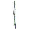
 Movie
Movie Controller


+ Open data

- Basic information
| Entry | Database: PDB / ID: 3nwh | |||||||||
|---|---|---|---|---|---|---|---|---|---|---|
| Title | Crystal structure of BST2/Tetherin | |||||||||
 Components Components | Bone marrow stromal antigen 2 | |||||||||
 Keywords Keywords |  ANTIVIRAL PROTEIN / Coiled-coil / innate immunity ANTIVIRAL PROTEIN / Coiled-coil / innate immunity | |||||||||
| Function / homology |  Function and homology information Function and homology informationnegative regulation of plasmacytoid dendritic cell cytokine production / negative regulation of intracellular transport of viral material / response to interferon-beta / response to interferon-alpha / metalloendopeptidase inhibitor activity / positive regulation of leukocyte proliferation / azurophil granule membrane / negative regulation of viral genome replication / B cell activation / response to type II interferon ...negative regulation of plasmacytoid dendritic cell cytokine production / negative regulation of intracellular transport of viral material / response to interferon-beta / response to interferon-alpha / metalloendopeptidase inhibitor activity / positive regulation of leukocyte proliferation / azurophil granule membrane / negative regulation of viral genome replication / B cell activation / response to type II interferon / side of membrane / multivesicular body / negative regulation of cell migration / regulation of actin cytoskeleton organization / response to virus / negative regulation of cell growth / SARS-CoV-1 activates/modulates innate immune responses / Interferon alpha/beta signaling / positive regulation of canonical NF-kappaB signal transduction / defense response to virus / membrane raft / apical plasma membrane / intracellular membrane-bounded organelle / innate immune response / Neutrophil degranulation / Golgi apparatus / cell surface / protein homodimerization activity / RNA binding / extracellular exosome / membrane / identical protein binding / plasma membraneSimilarity search - Function | |||||||||
| Biological species |  Homo sapiens (human) Homo sapiens (human) | |||||||||
| Method | X-RAY DIFFRACTION / SYNCHROTRON / MAD / Resolution: 2.6 Å | |||||||||
 Authors Authors | Schubert, H.L. / Zhai, Q. / Hill, C.P. | |||||||||
 Citation Citation | Journal: Proc.Natl.Acad.Sci.USA / Year: 2010 Title: Structural and functional studies on the extracellular domain of BST2/tetherin in reduced and oxidized conformations. Authors: Schubert, H.L. / Zhai, Q. / Sandrin, V. / Eckert, D.M. / Garcia-Maya, M. / Saul, L. / Sundquist, W.I. / Steiner, R.A. / Hill, C.P. | |||||||||
| History |
|
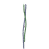
- Structure visualization
| Structure viewer | Molecule: MolmilJmol/JSmol |
|---|
- Downloads & links
-Download
| PDBx/mmCIF format | 3nwh.cif.gz | 171.2 KB | Display | PDBx/mmCIF format |
|---|---|---|---|---|
| PDB format | pdb3nwh.ent.gz | 145.6 KB | Display | PDB format |
| PDBx/mmJSON format | 3nwh.json.gz | Tree view | PDBx/mmJSON format | |
| Others |  Other downloads Other downloads |
-Validation report
| Arichive directory | https://data.pdbj.org/pub/pdb/validation_reports/nw/3nwhftp://data.pdbj.org/pub/pdb/validation_reports/nw/3nwh | HTTPS FTP |
|---|
-Related structure data


-Links
 PDBj
PDBj- Assembly
| Deposited unit | 
| ||||||||
|---|---|---|---|---|---|---|---|---|---|
| 1 |
| ||||||||
| Unit cell |
|
-Components
| #1: Protein | Mass: 12607.732 Da / Num. of mol.: 4 / Fragment: Extracellular Domain (UNP residues 47-152) Source method: isolated from a genetically manipulated source Source: (gene. exp.) Homo sapiens (human) / Gene: BST2 / Plasmid: pTOPO / Production host:  Escherichia coli (E. coli) / Strain (production host): BL21(DE3)RIL / References: UniProt: Q10589 Escherichia coli (E. coli) / Strain (production host): BL21(DE3)RIL / References: UniProt: Q10589#2: Water | ChemComp-HOH / | Water Mass: 18.015 Da / Num. of mol.: 18 / Source method: isolated from a natural source / Formula: H2O Mass: 18.015 Da / Num. of mol.: 18 / Source method: isolated from a natural source / Formula: H2O |
|---|
-Experimental details
-Experiment
| Experiment | Method: X-RAY DIFFRACTION / Number of used crystals: 1 |
|---|
- Sample preparation
| Crystal | Density Matthews: 2.5 Å3/Da / Density % sol: 50.81 % |
|---|---|
| Crystal grow | Temperature: 298 K / Method: vapor diffusion / pH: 7.2 Details: PEG MME 2K, pH 7.2, vapor diffusion, temperature 298K |
-Data collection
| Diffraction | Mean temperature: 100 K | |||||||||||||||||||||||||||||||||||||||||||||||||||||||||||||||||||||||||||||
|---|---|---|---|---|---|---|---|---|---|---|---|---|---|---|---|---|---|---|---|---|---|---|---|---|---|---|---|---|---|---|---|---|---|---|---|---|---|---|---|---|---|---|---|---|---|---|---|---|---|---|---|---|---|---|---|---|---|---|---|---|---|---|---|---|---|---|---|---|---|---|---|---|---|---|---|---|---|---|
| Diffraction source | Source: SYNCHROTRON / Site: SSRL  / Beamline: BL7-1 / Wavelength: 0.9805, 0.9919, 0.9809 / Beamline: BL7-1 / Wavelength: 0.9805, 0.9919, 0.9809 | |||||||||||||||||||||||||||||||||||||||||||||||||||||||||||||||||||||||||||||
| Detector | Type: ADSC QUANTUM 315r / Detector: CCD / Date: Dec 2, 2009 | |||||||||||||||||||||||||||||||||||||||||||||||||||||||||||||||||||||||||||||
| Radiation | Monochromator: side scattering I-beam bent single crystal; asymmetric cut 4.9650 deg Protocol: MAD / Monochromatic (M) / Laue (L): M / Scattering type: x-ray | |||||||||||||||||||||||||||||||||||||||||||||||||||||||||||||||||||||||||||||
| Radiation wavelength |
| |||||||||||||||||||||||||||||||||||||||||||||||||||||||||||||||||||||||||||||
| Reflection | Redundancy: 3.6 % / Av σ(I) over netI: 17.18 / Number: 15493 / Rmerge(I) obs: 0.058 / Χ2: 1.05 / D res high: 2.6 Å / D res low: 50 Å / Num. obs: 15096 / % possible obs: 97.5 | |||||||||||||||||||||||||||||||||||||||||||||||||||||||||||||||||||||||||||||
| Diffraction reflection shell |
| |||||||||||||||||||||||||||||||||||||||||||||||||||||||||||||||||||||||||||||
| Reflection | Resolution: 2.6→50 Å / Num. all: 15493 / Num. obs: 15096 / % possible obs: 97.5 % / Observed criterion σ(F): 0 / Observed criterion σ(I): 0 / Redundancy: 3.6 % / Rmerge(I) obs: 0.058 / Χ2: 1.048 / Net I/σ(I): 13 | |||||||||||||||||||||||||||||||||||||||||||||||||||||||||||||||||||||||||||||
| Reflection shell |
|
-Phasing
| Phasing | Method: MAD |
|---|
- Processing
| Software |
| |||||||||||||||||||||||||||||||||||||||||||||||||||||||||||||||||||||||||||||||||||||||||||||||||||||||||||||||||||||||||||||
|---|---|---|---|---|---|---|---|---|---|---|---|---|---|---|---|---|---|---|---|---|---|---|---|---|---|---|---|---|---|---|---|---|---|---|---|---|---|---|---|---|---|---|---|---|---|---|---|---|---|---|---|---|---|---|---|---|---|---|---|---|---|---|---|---|---|---|---|---|---|---|---|---|---|---|---|---|---|---|---|---|---|---|---|---|---|---|---|---|---|---|---|---|---|---|---|---|---|---|---|---|---|---|---|---|---|---|---|---|---|---|---|---|---|---|---|---|---|---|---|---|---|---|---|---|---|---|
| Refinement | Method to determine structure: MAD / Resolution: 2.6→33.13 Å / Cor.coef. Fo:Fc: 0.928 / Cor.coef. Fo:Fc free: 0.916 / Occupancy max: 1 / Occupancy min: 0.5 / SU B: 28.516 / SU ML: 0.275 / Cross valid method: THROUGHOUT / σ(F): 0 / ESU R: 1.346 / ESU R Free: 0.375 / Stereochemistry target values: MAXIMUM LIKELIHOOD / Details: HYDROGENS HAVE BEEN USED IF PRESENT IN THE INPUT
| |||||||||||||||||||||||||||||||||||||||||||||||||||||||||||||||||||||||||||||||||||||||||||||||||||||||||||||||||||||||||||||
| Solvent computation | Ion probe radii: 0.8 Å / Shrinkage radii: 0.8 Å / VDW probe radii: 1.2 Å / Solvent model: MASK | |||||||||||||||||||||||||||||||||||||||||||||||||||||||||||||||||||||||||||||||||||||||||||||||||||||||||||||||||||||||||||||
| Displacement parameters | Biso max: 161.21 Å2 / Biso mean: 56.2268 Å2 / Biso min: 20.2 Å2
| |||||||||||||||||||||||||||||||||||||||||||||||||||||||||||||||||||||||||||||||||||||||||||||||||||||||||||||||||||||||||||||
| Refinement step | Cycle: LAST / Resolution: 2.6→33.13 Å
| |||||||||||||||||||||||||||||||||||||||||||||||||||||||||||||||||||||||||||||||||||||||||||||||||||||||||||||||||||||||||||||
| Refine LS restraints |
| |||||||||||||||||||||||||||||||||||||||||||||||||||||||||||||||||||||||||||||||||||||||||||||||||||||||||||||||||||||||||||||
| LS refinement shell | Resolution: 2.6→2.667 Å / Total num. of bins used: 20
| |||||||||||||||||||||||||||||||||||||||||||||||||||||||||||||||||||||||||||||||||||||||||||||||||||||||||||||||||||||||||||||
| Refinement TLS params. | Method: refined / Refine-ID: X-RAY DIFFRACTION
| |||||||||||||||||||||||||||||||||||||||||||||||||||||||||||||||||||||||||||||||||||||||||||||||||||||||||||||||||||||||||||||
| Refinement TLS group |
|
