 Movie
Movie Controller

[English] 日本語
 Yorodumi
- PDB-3nt5: Crystal structure of myo-inositol dehydrogenase from Bacillus sub... -
+ Open data

- Basic information
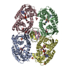
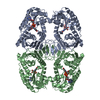
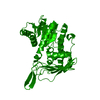
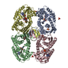
| Entry | Database: PDB / ID: 3nt5 | ||||||
|---|---|---|---|---|---|---|---|
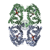
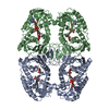
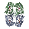
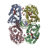
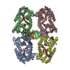
| Title | Crystal structure of myo-inositol dehydrogenase from Bacillus subtilis with bound cofactor and product inosose | ||||||
 Components Components | Inositol 2-dehydrogenase/D-chiro-inositol 3-dehydrogenase | ||||||
 Keywords Keywords |  OXIDOREDUCTASE / BSIDH / N-terminal Rossmann fold domain / glyceraldehyde-3-phosphate like C-terminal domain / NAD(H) / inositol / inosose OXIDOREDUCTASE / BSIDH / N-terminal Rossmann fold domain / glyceraldehyde-3-phosphate like C-terminal domain / NAD(H) / inositol / inosose | ||||||
| Function / homology |  Function and homology information Function and homology informationD-chiro-inositol 1-dehydrogenase / inositol 2-dehydrogenase / inositol 2-dehydrogenase (NAD+) activity / inositol catabolic process / nucleotide bindingSimilarity search - Function | ||||||
| Biological species |  Bacillus subtilis (bacteria) Bacillus subtilis (bacteria) | ||||||
| Method | X-RAY DIFFRACTION / SYNCHROTRON / MOLECULAR REPLACEMENT / molecular replacement / Resolution: 2.9006 Å | ||||||
 Authors Authors | Van Straaten, K.E. / Palmer, D.R.J. / Sanders, D.A.R. | ||||||
 Citation Citation | Journal: Biochem.J. / Year: 2010 Title: Structural investigation of myo-inositol dehydrogenase from Bacillus subtilis: implications for catalytic mechanism and inositol dehydrogenase subfamily classification. Authors: van Straaten, K.E. / Zheng, H. / Palmer, D.R. / Sanders, D.A. | ||||||
| History |
|
- Structure visualization
| Structure viewer | Molecule: MolmilJmol/JSmol |
|---|
- Downloads & links
-Download
| PDBx/mmCIF format | 3nt5.cif.gz | 134.8 KB | Display | PDBx/mmCIF format |
|---|---|---|---|---|
| PDB format | pdb3nt5.ent.gz | 106.7 KB | Display | PDB format |
| PDBx/mmJSON format | 3nt5.json.gz | Tree view | PDBx/mmJSON format | |
| Others |  Other downloads Other downloads |
-Validation report
| Arichive directory | https://data.pdbj.org/pub/pdb/validation_reports/nt/3nt5ftp://data.pdbj.org/pub/pdb/validation_reports/nt/3nt5 | HTTPS FTP |
|---|
-Related structure data
| Related structure data |  3mz0SC  3nt2C  3nt4C  3ntoC  3ntqC  3ntrC S: Starting model for refinement C: citing same article ( |
|---|---|
| Similar structure data |

-Links
 PDBj
PDBj

- Assembly
| Deposited unit | 
| |||||||||||||||||||||||||||||||||||||||||||||||||||||||||||||||||||||||||||||||
|---|---|---|---|---|---|---|---|---|---|---|---|---|---|---|---|---|---|---|---|---|---|---|---|---|---|---|---|---|---|---|---|---|---|---|---|---|---|---|---|---|---|---|---|---|---|---|---|---|---|---|---|---|---|---|---|---|---|---|---|---|---|---|---|---|---|---|---|---|---|---|---|---|---|---|---|---|---|---|---|---|
| 1 | 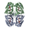
| |||||||||||||||||||||||||||||||||||||||||||||||||||||||||||||||||||||||||||||||
| 2 | 
| |||||||||||||||||||||||||||||||||||||||||||||||||||||||||||||||||||||||||||||||
| Unit cell |
| |||||||||||||||||||||||||||||||||||||||||||||||||||||||||||||||||||||||||||||||
| Noncrystallographic symmetry (NCS) | NCS domain:
NCS domain segments:
|