 Movie
Movie Controller

[English] 日本語
 Yorodumi
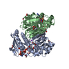
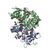
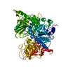
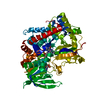
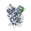
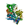
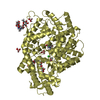
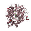
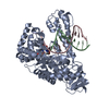
- PDB-3npf: Crystal structure of a putative dipeptidyl-peptidase VI (BACOVA_0... -
+ Open data

- Basic information
| Entry | Database: PDB / ID: 3npf | ||||||
|---|---|---|---|---|---|---|---|
| Title | Crystal structure of a putative dipeptidyl-peptidase VI (BACOVA_00612) from Bacteroides ovatus at 1.72 A resolution | ||||||
 Components Components | putative dipeptidyl-peptidase VI | ||||||
 Keywords Keywords |  HYDROLASE / Structural Genomics / Joint Center for Structural Genomics / JCSG / Protein Structure Initiative / PSI-2 HYDROLASE / Structural Genomics / Joint Center for Structural Genomics / JCSG / Protein Structure Initiative / PSI-2 | ||||||
| Function / homology |  Function and homology information Function and homology informationBacterial dipeptidyl-peptidase SH3 domain / Bacterial dipeptidyl-peptidase Sh3 domain / Endopeptidase, NLPC/P60 domain / NlpC/P60 family / endopeptidase domain like (from Nostoc punctiforme) / endopeptidase fold (from Nostoc punctiforme) / SH3 Domains / Papain-like cysteine peptidase superfamily / SH3 type barrels. / Roll ...Bacterial dipeptidyl-peptidase SH3 domain / Bacterial dipeptidyl-peptidase Sh3 domain / Endopeptidase, NLPC/P60 domain / NlpC/P60 family / endopeptidase domain like (from Nostoc punctiforme) / endopeptidase fold (from Nostoc punctiforme) / SH3 Domains / Papain-like cysteine peptidase superfamily / SH3 type barrels. / Roll / Alpha-Beta Complex / Mainly Beta / Alpha BetaSimilarity search - Domain/homology | ||||||
| Biological species |  Bacteroides ovatus (bacteria) Bacteroides ovatus (bacteria) | ||||||
| Method | X-RAY DIFFRACTION / SYNCHROTRON / SAD / Resolution: 1.72 Å | ||||||
 Authors Authors | Joint Center for Structural Genomics (JCSG) | ||||||
 Citation Citation | Journal: To be published Title: Crystal structure of a putative dipeptidyl-peptidase VI (BACOVA_00612) from Bacteroides ovatus at 1.72 A resolution Authors: Joint Center for Structural Genomics (JCSG) | ||||||
| History |
|
- Structure visualization
| Structure viewer | Molecule: MolmilJmol/JSmol |
|---|
- Downloads & links
-Download
| PDBx/mmCIF format | 3npf.cif.gz | 262.5 KB | Display | PDBx/mmCIF format |
|---|---|---|---|---|
| PDB format | pdb3npf.ent.gz | 215.4 KB | Display | PDB format |
| PDBx/mmJSON format | 3npf.json.gz | Tree view | PDBx/mmJSON format | |
| Others |  Other downloads Other downloads |
-Validation report
| Arichive directory | https://data.pdbj.org/pub/pdb/validation_reports/np/3npfftp://data.pdbj.org/pub/pdb/validation_reports/np/3npf | HTTPS FTP |
|---|
-Related structure data
| Similar structure data | |
|---|---|
| Other databases |
-Links
 PDBj
PDBj- Assembly
| Deposited unit | 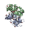
| |||||||||||||||||||||||||||
|---|---|---|---|---|---|---|---|---|---|---|---|---|---|---|---|---|---|---|---|---|---|---|---|---|---|---|---|---|
| 1 |
| |||||||||||||||||||||||||||
| Unit cell |
| |||||||||||||||||||||||||||
| Noncrystallographic symmetry (NCS) | NCS domain:
NCS domain segments:
|
-Components
| #1: Protein | Mass: 35155.070 Da / Num. of mol.: 2 Source method: isolated from a genetically manipulated source Source: (gene. exp.) Bacteroides ovatus (bacteria) / Strain: ATCC 8483 / Gene: BACOVA_00612 / Plasmid: SpeedET / Production host: Escherichia Coli (E. coli) / Strain (production host): HK100 / References: UniProt: A7LS31#2: Chemical | Chloride  Mass: 35.453 Da / Num. of mol.: 2 / Source method: obtained synthetically / Formula: Cl Mass: 35.453 Da / Num. of mol.: 2 / Source method: obtained synthetically / Formula: Cl#3: Chemical | ChemComp-GOL / Glycerol  Mass: 92.094 Da / Num. of mol.: 11 / Source method: obtained synthetically / Formula: C3H8O3 Mass: 92.094 Da / Num. of mol.: 11 / Source method: obtained synthetically / Formula: C3H8O3#4: Water | ChemComp-HOH / | Water Mass: 18.015 Da / Num. of mol.: 786 / Source method: isolated from a natural source / Formula: H2O Mass: 18.015 Da / Num. of mol.: 786 / Source method: isolated from a natural source / Formula: H2OSequence details | THE CONSTRUCT (RESIDUES 22-326) WAS EXPRESSED WITH A PURIFICATION TAG MGSDKIHHHHHHENLYFQG. THE TAG ...THE CONSTRUCT (RESIDUES 22-326) WAS EXPRESSED WITH A PURIFICATI | |
|---|
-Experimental details
-Experiment
| Experiment | Method: X-RAY DIFFRACTION / Number of used crystals: 1 |
|---|
- Sample preparation
| Crystal | Density Matthews: 2.1 Å3/Da / Density % sol: 41.3 % |
|---|---|
| Crystal grow | Temperature: 277 K / Method: vapor diffusion, sitting drop / pH: 8.5 Details: 15.000000000% Glycerol, 0.170000000M NaOAc, 25.500000000% PEG-4000, 0.1M TRIS pH 8.5, NANODROP', VAPOR DIFFUSION, SITTING DROP, temperature 277K |
-Data collection
| Diffraction | Mean temperature: 100 K | ||||||||||||||||||||||||||||||||||||||||||||||||||||||||||||||||||
|---|---|---|---|---|---|---|---|---|---|---|---|---|---|---|---|---|---|---|---|---|---|---|---|---|---|---|---|---|---|---|---|---|---|---|---|---|---|---|---|---|---|---|---|---|---|---|---|---|---|---|---|---|---|---|---|---|---|---|---|---|---|---|---|---|---|---|---|
| Diffraction source | Source: SYNCHROTRON / Site: SSRL  / Beamline: BL9-2 / Wavelength: 0.97925 / Beamline: BL9-2 / Wavelength: 0.97925 | ||||||||||||||||||||||||||||||||||||||||||||||||||||||||||||||||||
| Detector | Type: MARMOSAIC 325 mm CCD / Detector: CCD / Date: May 13, 2010 / Details: Flat collimating mirror, toroid focusing mirror | ||||||||||||||||||||||||||||||||||||||||||||||||||||||||||||||||||
| Radiation | Monochromator: Double crystal monochromator / Protocol: SINGLE WAVELENGTH / Monochromatic (M) / Laue (L): M / Scattering type: x-ray | ||||||||||||||||||||||||||||||||||||||||||||||||||||||||||||||||||
| Radiation wavelength | Wavelength: 0.97925 Å / Relative weight: 1 | ||||||||||||||||||||||||||||||||||||||||||||||||||||||||||||||||||
| Reflection | Resolution: 1.72→48.87 Å / Num. obs: 63712 / % possible obs: 100 % / Observed criterion σ(I): -3 / Redundancy: 6.96 % / Biso Wilson estimate: 16.41 Å2 / Rmerge(I) obs: 0.125 / Net I/σ(I): 11.06 | ||||||||||||||||||||||||||||||||||||||||||||||||||||||||||||||||||
| Reflection shell |
|
-Phasing
| Phasing | Method: SAD |
|---|
- Processing
| Software |
| |||||||||||||||||||||||||||||||||||||||||||||||||||||||||||||||||||||||||||||||||||||
|---|---|---|---|---|---|---|---|---|---|---|---|---|---|---|---|---|---|---|---|---|---|---|---|---|---|---|---|---|---|---|---|---|---|---|---|---|---|---|---|---|---|---|---|---|---|---|---|---|---|---|---|---|---|---|---|---|---|---|---|---|---|---|---|---|---|---|---|---|---|---|---|---|---|---|---|---|---|---|---|---|---|---|---|---|---|---|
| Refinement | Method to determine structure: SAD / Resolution: 1.72→48.87 Å / Cor.coef. Fo:Fc: 0.972 / Cor.coef. Fo:Fc free: 0.952 / Occupancy max: 1 / Occupancy min: 0.25 / SU B: 3.676 / SU ML: 0.061 / Cross valid method: THROUGHOUT / σ(F): 0 / ESU R Free: 0.092 Stereochemistry target values: MAXIMUM LIKELIHOOD WITH PHASES Details: 1. HYDROGENS HAVE BEEN ADDED IN THE RIDING POSITIONS. 2. A MET-INHIBITION PROTOCOL WAS USED FOR SELENOMETHIONINE INCORPORATION DURING PROTEIN EXPRESSION. THE OCCUPANCY OF THE SE ATOMS IN THE ...Details: 1. HYDROGENS HAVE BEEN ADDED IN THE RIDING POSITIONS. 2. A MET-INHIBITION PROTOCOL WAS USED FOR SELENOMETHIONINE INCORPORATION DURING PROTEIN EXPRESSION. THE OCCUPANCY OF THE SE ATOMS IN THE MSE RESIDUES WAS REDUCED TO 0.75 FOR THE REDUCED SCATTERING POWER DUE TO PARTIAL S-MET INCORPORATION. 3. ATOM RECORD CONTAINS SUM OF TLS AND RESIDUAL B FACTORS. ANISOU RECORD CONTAINS SUM OF TLS AND RESIDUAL U FACTORS. 4. WATERS WERE EXCLUDED FROM AUTOMATIC TLS ASSIGNMENT. 5. GLYCEROL (GOL) AND CHLORIDE (CL) MODELED ARE PRESENT IN CRYSTALLIZATION/CRYO CONDITIONS. 6. ACTIVE SITE RESIDUE CYS203 IS COVALENTLY MODIFIED. IT IS MODELED AS S-ACETONYLCYSTEINE (CSA) BASED ON DENSITY AND INTERACTION ENVIROMENT.
| |||||||||||||||||||||||||||||||||||||||||||||||||||||||||||||||||||||||||||||||||||||
| Solvent computation | Ion probe radii: 0.8 Å / Shrinkage radii: 0.8 Å / VDW probe radii: 1.2 Å / Solvent model: MASK | |||||||||||||||||||||||||||||||||||||||||||||||||||||||||||||||||||||||||||||||||||||
| Displacement parameters | Biso max: 71.14 Å2 / Biso mean: 18.5442 Å2 / Biso min: 5.48 Å2
| |||||||||||||||||||||||||||||||||||||||||||||||||||||||||||||||||||||||||||||||||||||
| Refinement step | Cycle: LAST / Resolution: 1.72→48.87 Å
| |||||||||||||||||||||||||||||||||||||||||||||||||||||||||||||||||||||||||||||||||||||
| Refine LS restraints |
| |||||||||||||||||||||||||||||||||||||||||||||||||||||||||||||||||||||||||||||||||||||
| Refine LS restraints NCS | Dom-ID: 1 / Auth asym-ID: A / Ens-ID: 1 / Refine-ID: X-RAY DIFFRACTION
| |||||||||||||||||||||||||||||||||||||||||||||||||||||||||||||||||||||||||||||||||||||
| LS refinement shell | Resolution: 1.72→1.765 Å / Total num. of bins used: 20
| |||||||||||||||||||||||||||||||||||||||||||||||||||||||||||||||||||||||||||||||||||||
| Refinement TLS params. | Method: refined / Refine-ID: X-RAY DIFFRACTION
| |||||||||||||||||||||||||||||||||||||||||||||||||||||||||||||||||||||||||||||||||||||
| Refinement TLS group |
|
