 Movie
Movie Controller

[English] 日本語
 Yorodumi
- PDB-3nb3: The host outer membrane proteins OmpA and OmpC are packed at spec... -
+ Open data

- Basic information
| Entry | Database: PDB / ID: 3nb3 | ||||||
|---|---|---|---|---|---|---|---|
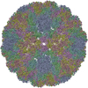
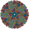
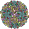
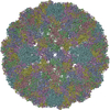
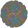
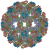
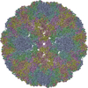
| Title | The host outer membrane proteins OmpA and OmpC are packed at specific sites in the Shigella phage Sf6 virion as structural components | ||||||
 Components Components |
| ||||||
 Keywords Keywords |  MEMBRANE PROTEIN / virus assembly / cementing protein / bacteriophage / Sf6 / Shigella / beta-barrel / outer membrane protein / icosahedral MEMBRANE PROTEIN / virus assembly / cementing protein / bacteriophage / Sf6 / Shigella / beta-barrel / outer membrane protein / icosahedral | ||||||
| Function / homology |  Function and homology information Function and homology informationouter membrane protein complex / monoatomic ion transmembrane transporter activity / detection of virus / outer membrane / porin activity / pore complex / monoatomic ion transport / monoatomic ion transmembrane transport / cell outer membrane / virus receptor activity ...outer membrane protein complex / monoatomic ion transmembrane transporter activity / detection of virus / outer membrane / porin activity / pore complex / monoatomic ion transport / monoatomic ion transmembrane transport / cell outer membrane / virus receptor activity / outer membrane-bounded periplasmic space / receptor-mediated virion attachment to host cell / symbiont entry into host cell / DNA damage response / membrane / identical protein binding / metal ion bindingSimilarity search - Function | ||||||
| Biological species |  Escherichia coli (E. coli) Escherichia coli (E. coli) | ||||||
| Method | ELECTRON MICROSCOPY / single particle reconstruction / cryo EM / Resolution: 19 Å | ||||||
 Authors Authors | Zhao, H. / Sequeira, R.D. / Galeva, N.A. / Tang, L. | ||||||
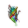
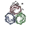
 Citation Citation | Journal: Virology / Year: 2011 Title: The host outer membrane proteins OmpA and OmpC are associated with the Shigella phage Sf6 virion. Authors: Haiyan Zhao / Reuben D Sequeira / Nadezhda A Galeva / Liang Tang /  Abstract: Assembly of dsDNA bacteriophage is a precisely programmed process. Potential roles of host cell components in phage assembly haven't been well understood. It was previously reported that two ...Assembly of dsDNA bacteriophage is a precisely programmed process. Potential roles of host cell components in phage assembly haven't been well understood. It was previously reported that two unidentified proteins were present in bacteriophage Sf6 virion (Casjens et al, 2004, J.Mol.Biol. 339, 379-394, Fig. 2A). Using tandem mass spectrometry, we have identified the two proteins as outer membrane proteins (OMPs) OmpA and OmpC from its host Shigella flexneri. The transmission electron cryo-microscopy structure of Sf6 shows significant density at specific sites at the phage capsid inner surface. This density fit well with the characteristic beta-barrel domains of OMPs, thus may be due to the two host proteins. Locations of this density suggest a role in Sf6 morphogenesis reminiscent of phage-encoded cementing proteins. These data indicate a new, OMP-related phage:host linkage, adding to previous knowledge that some lambdoid bacteriophage genomes contain OmpC-like genes that express phage-encoded porins in the lysogenic state. | ||||||
| History |
|
- Structure visualization
| Movie |
Movie viewer |
|---|---|
| Structure viewer | Molecule: MolmilJmol/JSmol |
- Downloads & links
-Download
| PDBx/mmCIF format | 3nb3.cif.gz | 159.5 KB | Display | PDBx/mmCIF format |
|---|---|---|---|---|
| PDB format | pdb3nb3.ent.gz | 119.4 KB | Display | PDB format |
| PDBx/mmJSON format | 3nb3.json.gz | Tree view | PDBx/mmJSON format | |
| Others |  Other downloads Other downloads |
-Validation report
| Arichive directory | https://data.pdbj.org/pub/pdb/validation_reports/nb/3nb3ftp://data.pdbj.org/pub/pdb/validation_reports/nb/3nb3 | HTTPS FTP |
|---|
-Related structure data
| Related structure data | 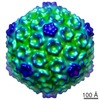 5201MC M: map data used to model this data C: citing same article ( |
|---|---|
| Similar structure data |

-Links
 PDBj
PDBj

- Assembly
| Deposited unit | 
|
|---|---|
| 1 | x 60
|
| 2 |
|
| 3 | x 5
|
| 4 | x 6
|
| 5 | 
|
| Symmetry | Point symmetry: (Schoenflies symbol: I (icosahedral)) |
| Details | CHAIN C IS LOCATED ON AN ICOSAHEDRAL 2-FOLD AXIS AND HAS 0.50 OCCUPANCY. CHAIN D IS LOCATED ON AN ICOSAHEDRAL 3-FOLD AXIS AND HAS 0.33 OCCUPANCY. |
-Components
| #1: Protein | Mass: 37272.672 Da / Num. of mol.: 3 / Fragment: outer membrane protein A / Source method: isolated from a natural source / Source: (natural) Escherichia coli (E. coli) / Strain: K12 / References: UniProt: P0A910#2: Protein | | Mass: 38336.242 Da / Num. of mol.: 1 / Fragment: outer membrane protein C / Source method: isolated from a natural source / Source: (natural) Escherichia coli (E. coli) / Strain: K12 / References: UniProt: P06996Sequence details | AUTHORS STATE THAT THE SEQUENCE CONFLICTS ARE INHERITED FROM IN THE ORIGINAL PDB ENTRY 1QJP USED FOR FITTING. | |
|---|
-Experimental details
-Experiment
| Experiment | Method: ELECTRON MICROSCOPY |
|---|---|
| EM experiment | Aggregation state: PARTICLE / 3D reconstruction method: single particle reconstruction |
- Sample preparation
| Component | Name: bacteriophage Sf6 / Type: VIRUS / Synonym: Sf6 |
|---|---|
| Molecular weight | Value: 19.2 MDa / Experimental value: NO |
| Details of virus | Host category: GRAM-NEGATIVE BACTERIA / Isolate: SPECIES / Type: VIRION |
| Natural host | Organism: Shigella flexneri |
| Virus shell | Name: gp5 / Diameter: 680 nm / Triangulation number (T number): 7 |
| Buffer solution | Name: 10 mM Tris-HCl, pH 7.4, 10 mM MgCl2 / pH: 7.4 / Details: 10 mM Tris-HCl, pH 7.4, 10 mM MgCl2 |
| Specimen | Conc.: 2 mg/ml / Embedding applied: NO / Shadowing applied: NO / Staining applied: NO / Vitrification applied: YES |
| Specimen support | Details: 300 mesh holey copper grids |
| Vitrification | Instrument: HOMEMADE PLUNGER / Cryogen name: ETHANE Details: Vitrification was carried out with a standard cryoEM method. The sample of 4 microlitres was applied to a holey EM grid, blotted for 3-4 seconds with a Whatman #2 filter paper, and plunged ...Details: Vitrification was carried out with a standard cryoEM method. The sample of 4 microlitres was applied to a holey EM grid, blotted for 3-4 seconds with a Whatman #2 filter paper, and plunged into ethane slush incubated in liquid nitrogen. The grid was stored in liquid nitrogen. |
- Electron microscopy imaging
| Experimental equipment |  Model: Tecnai F20 / Image courtesy: FEI Company |
|---|---|
| Microscopy | Model: FEI TECNAI F20 / Date: Jul 18, 2008 |
| Electron gun | Electron source: FIELD EMISSION GUN / Accelerating voltage: 200 kV / Illumination mode: FLOOD BEAM |
| Electron lens | Mode: BRIGHT FIELDBright-field microscopy / Nominal magnification: 39000 X / Nominal defocus max: 471 nm / Nominal defocus min: 1060 nm / Cs: 4 mm |
| Specimen holder | Temperature: 89 K / Tilt angle max: 0 ° / Tilt angle min: 0 ° |
| Image recording | Electron dose: 20 e/Å2 / Film or detector model: GENERIC CCD (2k x 2k) |
- Processing
| CTF correction | Details: none | |||||||||||||||||||||
|---|---|---|---|---|---|---|---|---|---|---|---|---|---|---|---|---|---|---|---|---|---|---|
| Symmetry | Point symmetry: I (icosahedral) | |||||||||||||||||||||
| 3D reconstruction | Method: model-based projection / Resolution: 19 Å / Num. of particles: 3232 / Nominal pixel size: 4.05 Å / Actual pixel size: 3.9284 Å / Magnification calibration: none Details: The initial model was the cryoEM map of the related phage P22. Symmetry type: POINT | |||||||||||||||||||||
| Atomic model building | Protocol: RIGID BODY FIT / Space: REAL Details: METHOD--manual fitting in O coupled with local optimization with UCSF Chimera REFINEMENT PROTOCOL--rigid body | |||||||||||||||||||||
| Atomic model building |
| |||||||||||||||||||||
| Refinement step | Cycle: LAST
|
