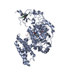
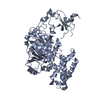
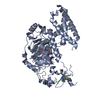
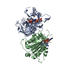
OXIDOREDUCTASE / Histone / methylation / demethylase / PHD / JmjC / Fe(II) and alpha-KG (alpha-ketoglutarate)-dependent dioxygenase family
Function / homology
Function and homology information
HDMs demethylate histones / Condensation of Prophase Chromosomes / histone H3K27me2/H3K27me3 demethylase activity / Oxidoreductases; Acting on paired donors, with incorporation or reduction of molecular oxygen; With 2-oxoglutarate as one donor, and incorporation of one atom of oxygen into each donor / histone H3K9 demethylase activity / histone demethylase activity / transcription coregulator activity / histone binding / regulation of transcription by RNA polymerase II / metal ion binding / nucleus Similarity search - Function
Methane Monooxygenase Hydroxylase; Chain G, domain 1 - #1360 / Jumonji, helical domain / Jumonji helical domain / Zinc/RING finger domain, C3HC4 (zinc finger) / Cupin / Herpes Virus-1 / JmjC domain, hydroxylase / A domain family that is part of the cupin metalloenzyme superfamily. / JmjC domain / JmjC domain profile. ...Methane Monooxygenase Hydroxylase; Chain G, domain 1 - #1360 / Jumonji, helical domain / Jumonji helical domain / Zinc/RING finger domain, C3HC4 (zinc finger) / Cupin / Herpes Virus-1 / JmjC domain, hydroxylase / A domain family that is part of the cupin metalloenzyme superfamily. / JmjC domain / JmjC domain profile. / Zinc finger, PHD-type, conserved site / Zinc finger PHD-type signature. / Methane Monooxygenase Hydroxylase; Chain G, domain 1 / Zinc finger, PHD-type / PHD zinc finger / Zinc finger, FYVE/PHD-type / Zinc finger, RING/FYVE/PHD-type / Jelly Rolls / Up-down Bundle / Sandwich / 2-Layer Sandwich / Mainly Beta / Mainly Alpha / Alpha Beta Similarity search - Domain/homology
In the structure databanks used in Yorodumi, some data are registered as the other names, "COVID-19 virus" and "2019-nCoV". Here are the details of the virus and the list of structure data.
Jan 31, 2019. EMDB accession codes are about to change! (news from PDBe EMDB page)
EMDB accession codes are about to change! (news from PDBe EMDB page)
The allocation of 4 digits for EMDB accession codes will soon come to an end. Whilst these codes will remain in use, new EMDB accession codes will include an additional digit and will expand incrementally as the available range of codes is exhausted. The current 4-digit format prefixed with “EMD-” (i.e. EMD-XXXX) will advance to a 5-digit format (i.e. EMD-XXXXX), and so on. It is currently estimated that the 4-digit codes will be depleted around Spring 2019, at which point the 5-digit format will come into force.
The EM Navigator/Yorodumi systems omit the EMD- prefix.
Related info.:Q: What is EMD? / ID/Accession-code notation in Yorodumi/EM Navigator
Yorodumi is a browser for structure data from EMDB, PDB, SASBDB, etc.
This page is also the successor to EM Navigator detail page, and also detail information page/front-end page for Omokage search.
The word "yorodu" (or yorozu) is an old Japanese word meaning "ten thousand". "mi" (miru) is to see.
Related info.:EMDB / PDB / SASBDB / Comparison of 3 databanks / Yorodumi Search / Aug 31, 2016. New EM Navigator & Yorodumi / Yorodumi Papers / Jmol/JSmol / Function and homology information / Changes in new EM Navigator and Yorodumi
 Movie
Movie Controller


 Open data
 Basic information
 Components
 Keywords
 OXIDOREDUCTASE /
  Function and homology information
 Authors
 Citation
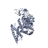
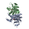
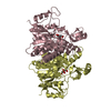
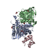
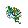
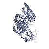
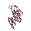
 Structure visualization
 Downloads & links
 Other downloads
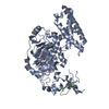


 PDBj
PDBj


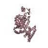
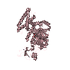
 Assembly


 Mass: 55.845 Da / Num. of mol.: 2 / Source method: obtained synthetically / Formula: Fe
 Mass: 65.409 Da / Num. of mol.: 4 / Source method: obtained synthetically / Formula: Zn
 Mass: 18.015 Da / Num. of mol.: 220 / Source method: isolated from a natural source / Formula: H2O
 Sample preparation
 / Beamline: BL-17A / Wavelength: 1 Å
 Processing