 Movie
Movie Controller

[English] 日本語
 Yorodumi
- PDB-3n7u: NAD-dependent formate dehydrogenase from higher-plant Arabidopsis... -
+ Open data

- Basic information
| Entry | Database: PDB / ID: 3n7u | ||||||
|---|---|---|---|---|---|---|---|
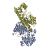
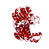
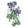
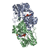
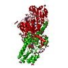
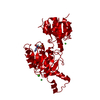
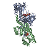
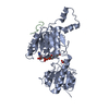
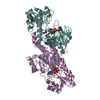
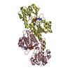
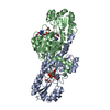
| Title | NAD-dependent formate dehydrogenase from higher-plant Arabidopsis thaliana in complex with NAD and azide | ||||||
 Components Components | Formate dehydrogenase | ||||||
 Keywords Keywords | OXIDOREDUCTASE / homodimer / Holo-form | ||||||
| Function / homology |  Function and homology information Function and homology informationformate catabolic process / formate dehydrogenase / formate dehydrogenase (NAD+) activity / thylakoid / oxidoreductase activity, acting on the CH-OH group of donors, NAD or NADP as acceptor / plastid / chloroplast / NAD binding / mitochondrion / cytosolSimilarity search - Function | ||||||
| Biological species |  Arabidopsis thaliana (thale cress) Arabidopsis thaliana (thale cress) | ||||||
| Method | X-RAY DIFFRACTION / SYNCHROTRON / MOLECULAR REPLACEMENT / molecular replacement / Resolution: 2 Å | ||||||
 Authors Authors | Shabalin, I.G. / Polyakov, K.M. / Serov, A.E. / Skirgello, O.E. / Sadykhov, E.G. / Dorovatovskiy, P.V. / Tishkov, V.I. / Popov, V.O. | ||||||
 Citation Citation | Journal: To be Published Title: Structures of the apo and holo forms of NAD-dependent formate dehydrogenase from the higher-plant Arabidopsis Thaliana Authors: Shabalin, I.G. / Polyakov, K.M. / Skirgello, O.E. / Tishkov, V.I. / Popov, V.O. | ||||||
| History |
|
- Structure visualization
| Structure viewer | Molecule: MolmilJmol/JSmol |
|---|
- Downloads & links
-Download
| PDBx/mmCIF format | 3n7u.cif.gz | 897.2 KB | Display | PDBx/mmCIF format |
|---|---|---|---|---|
| PDB format | pdb3n7u.ent.gz | 744.5 KB | Display | PDB format |
| PDBx/mmJSON format | 3n7u.json.gz | Tree view | PDBx/mmJSON format | |
| Others |  Other downloads Other downloads |
-Validation report
| Arichive directory | https://data.pdbj.org/pub/pdb/validation_reports/n7/3n7uftp://data.pdbj.org/pub/pdb/validation_reports/n7/3n7u | HTTPS FTP |
|---|
-Related structure data


-Links
 PDBj
PDBj- Assembly
| Deposited unit | 
| ||||||||
|---|---|---|---|---|---|---|---|---|---|
| 1 | 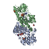
| ||||||||
| 2 | 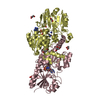
| ||||||||
| 3 | 
| ||||||||
| 4 | 
| ||||||||
| 5 | 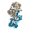
| ||||||||
| 6 | 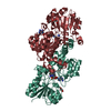
| ||||||||
| Unit cell |
|
-Components
-Protein , 1 types, 12 molecules ABCDEFGHIJKL
| #1: Protein | / NAD-dependent formate dehydrogenase / FDH Mass: 38990.621 Da / Num. of mol.: 12 Source method: isolated from a genetically manipulated source Source: (gene. exp.) Arabidopsis thaliana (thale cress) / Gene: At5g14780, FDH, FDH1, T9L3_80 / Plasmid: p1AraFDH / Production host:  Escherichia coli (E. coli) / Strain (production host): Bl21(DE3) / References: UniProt: Q9S7E4, formate dehydrogenase Escherichia coli (E. coli) / Strain (production host): Bl21(DE3) / References: UniProt: Q9S7E4, formate dehydrogenase |
|---|
-Non-polymers , 5 types, 3797 molecules 


| #2: Chemical | ChemComp-NAD / Nicotinamide adenine dinucleotide Mass: 663.425 Da / Num. of mol.: 12 / Source method: obtained synthetically / Formula: C21H27N7O14P2 / Comment: NAD*YM Mass: 663.425 Da / Num. of mol.: 12 / Source method: obtained synthetically / Formula: C21H27N7O14P2 / Comment: NAD*YM#3: Chemical | ChemComp-AZI / Azide Mass: 42.020 Da / Num. of mol.: 12 / Source method: obtained synthetically / Formula: N3 Mass: 42.020 Da / Num. of mol.: 12 / Source method: obtained synthetically / Formula: N3#4: Chemical | ChemComp-SO4 / Sulfate Mass: 96.063 Da / Num. of mol.: 9 / Source method: obtained synthetically / Formula: SO4 Mass: 96.063 Da / Num. of mol.: 9 / Source method: obtained synthetically / Formula: SO4#5: Chemical | ChemComp-GOL / Glycerol Mass: 92.094 Da / Num. of mol.: 19 / Source method: obtained synthetically / Formula: C3H8O3 Mass: 92.094 Da / Num. of mol.: 19 / Source method: obtained synthetically / Formula: C3H8O3#6: Water | ChemComp-HOH / | WaterMass: 18.015 Da / Num. of mol.: 3745 / Source method: isolated from a natural source / Formula: H2O |
|---|
-Experimental details
-Experiment
| Experiment | Method: X-RAY DIFFRACTION / Number of used crystals: 1 |
|---|
- Sample preparation
| Crystal | Density Matthews: 3.71 Å3/Da / Density % sol: 66.86 % |
|---|---|
| Crystal grow | Temperature: 293 K / Method: vapor diffusion, hanging drop Details: Protein solution (2ml): 14 mg/ml FDH, 0.1M Na2HPO4, pH 7.0, 10 mM EDTA, 5mM sodium azide, 5mM NAD. Reservoir solution (2ml): 0.1M Bis-Tris, pH 5.5, 2.1M Ammonium Sulfate, VAPOR DIFFUSION, ...Details: Protein solution (2ml): 14 mg/ml FDH, 0.1M Na2HPO4, pH 7.0, 10 mM EDTA, 5mM sodium azide, 5mM NAD. Reservoir solution (2ml): 0.1M Bis-Tris, pH 5.5, 2.1M Ammonium Sulfate, VAPOR DIFFUSION, HANGING DROP, temperature 293K |
-Data collection
| Diffraction | Mean temperature: 100 K |
|---|---|
| Diffraction source | Source: SYNCHROTRON / Site: KURCHATOV SNC  / Beamline: K4.4 / Wavelength: 0.99 Å / Beamline: K4.4 / Wavelength: 0.99 Å |
| Detector | Type: MAR CCD 165 mm / Detector: CCD |
| Radiation | Protocol: SINGLE WAVELENGTH / Monochromatic (M) / Laue (L): M / Scattering type: x-ray |
| Radiation wavelength | Wavelength: 0.99 Å / Relative weight: 1 |
| Reflection | Resolution: 2→19.99 Å / Num. all: 457766 / Num. obs: 428654 / % possible obs: 93.6 % / Observed criterion σ(F): 0 / Observed criterion σ(I): 0 / Redundancy: 2.1 % / Biso Wilson estimate: 25.095 Å2 / Rmerge(I) obs: 0.075 / Net I/σ(I): 8.93 |
| Reflection shell | Resolution: 2→2.07 Å / Redundancy: 1.8 % / Rmerge(I) obs: 0.328 / Mean I/σ(I) obs: 2.6 / Num. measured obs: 75579 / Num. unique all: 39349 / Num. unique obs: 42823 / % possible all: 87.9 |
-Phasing
| Phasing | Method: molecular replacement |
|---|
- Processing
| Software |
| |||||||||||||||||||||||||||||||||||||||||||||||||||||||||||||||||
|---|---|---|---|---|---|---|---|---|---|---|---|---|---|---|---|---|---|---|---|---|---|---|---|---|---|---|---|---|---|---|---|---|---|---|---|---|---|---|---|---|---|---|---|---|---|---|---|---|---|---|---|---|---|---|---|---|---|---|---|---|---|---|---|---|---|---|
| Refinement | Method to determine structure: MOLECULAR REPLACEMENT / Resolution: 2→19.99 Å / Cor.coef. Fo:Fc: 0.953 / Cor.coef. Fo:Fc free: 0.928 / WRfactor Rfree: 0.206 / WRfactor Rwork: 0.168 / Occupancy max: 1 / Occupancy min: 0.5 / FOM work R set: 0.839 / SU B: 3.551 / SU ML: 0.097 / SU R Cruickshank DPI: 0.144 / SU Rfree: 0.141 / Cross valid method: THROUGHOUT / σ(F): 0 / ESU R Free: 0.141 / Stereochemistry target values: MAXIMUM LIKELIHOOD / Details: U VALUES REFINED INDIVIDUALLY
| |||||||||||||||||||||||||||||||||||||||||||||||||||||||||||||||||
| Solvent computation | Ion probe radii: 0.8 Å / Shrinkage radii: 0.8 Å / VDW probe radii: 1.4 Å / Solvent model: BABINET MODEL WITH MASK | |||||||||||||||||||||||||||||||||||||||||||||||||||||||||||||||||
| Displacement parameters | Biso max: 62.55 Å2 / Biso mean: 24.463 Å2 / Biso min: 6.15 Å2
| |||||||||||||||||||||||||||||||||||||||||||||||||||||||||||||||||
| Refine analyze | Luzzati coordinate error obs: 0.226 Å | |||||||||||||||||||||||||||||||||||||||||||||||||||||||||||||||||
| Refinement step | Cycle: LAST / Resolution: 2→19.99 Å
| |||||||||||||||||||||||||||||||||||||||||||||||||||||||||||||||||
| Refine LS restraints |
| |||||||||||||||||||||||||||||||||||||||||||||||||||||||||||||||||
| LS refinement shell | Resolution: 2→2.051 Å / Total num. of bins used: 20
|
