 Movie
Movie Controller

[English] 日本語
 Yorodumi
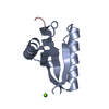
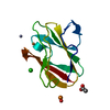
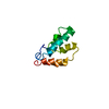
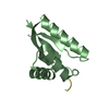
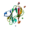
- PDB-3n4z: Crystal structure of quintuple Arg-to-Lys variant of T. celer L30e -
+ Open data

- Basic information
| Entry | Database: PDB / ID: 3n4z | ||||||
|---|---|---|---|---|---|---|---|
| Title | Crystal structure of quintuple Arg-to-Lys variant of T. celer L30e | ||||||
 Components Components | 50S ribosomal protein L30e Ribosome Ribosome | ||||||
 Keywords Keywords | RIBOSOMAL PROTEIN / L30e / quintuple / Arg-to-Lys | ||||||
| Function / homology |  Function and homology information Function and homology informationcytosolic large ribosomal subunit / structural constituent of ribosome / translation / RNA bindingSimilarity search - Function | ||||||
| Biological species |   Thermococcus celer (archaea) Thermococcus celer (archaea) | ||||||
| Method | X-RAY DIFFRACTION / MOLECULAR REPLACEMENT / Resolution: 2.3973 Å | ||||||
 Authors Authors | Chan, C.H. / Wong, K.B. | ||||||
 Citation Citation | Journal: Plos One / Year: 2012 Title: Electrostatic contribution of surface charge residues to the stability of a thermophilic protein: benchmarking experimental and predicted pKa values Authors: Chan, C.H. / Wilbanks, C.C. / Makhatadze, G.I. / Wong, K.B. | ||||||
| History |
|
- Structure visualization
| Structure viewer | Molecule: MolmilJmol/JSmol |
|---|
- Downloads & links
-Download
| PDBx/mmCIF format | 3n4z.cif.gz | 50.2 KB | Display | PDBx/mmCIF format |
|---|---|---|---|---|
| PDB format | pdb3n4z.ent.gz | 35.5 KB | Display | PDB format |
| PDBx/mmJSON format | 3n4z.json.gz | Tree view | PDBx/mmJSON format | |
| Others |  Other downloads Other downloads |
-Validation report
| Arichive directory | https://data.pdbj.org/pub/pdb/validation_reports/n4/3n4zftp://data.pdbj.org/pub/pdb/validation_reports/n4/3n4z | HTTPS FTP |
|---|
-Related structure data
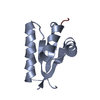
| Related structure data | 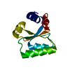 3n4yC 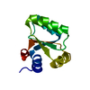 1h7mS S: Starting model for refinement C: citing same article ( |
|---|---|
| Similar structure data |


-Links
 PDBj
PDBj


- Assembly
| Deposited unit | 
| ||||||||
|---|---|---|---|---|---|---|---|---|---|
| 1 | 
| ||||||||
| 2 | 
| ||||||||
| Unit cell |
|
-Components
| #1: Protein | Ribosome Mass: 10841.639 Da / Num. of mol.: 2 / Mutation: R8K, R21K, R42K, R54K, R76K Source method: isolated from a genetically manipulated source Source: (gene. exp.) Thermococcus celer (archaea) / Plasmid: pET3C / Production host:  Escherichia coli (E. coli) / Strain (production host): C41 / References: UniProt: P29160 Escherichia coli (E. coli) / Strain (production host): C41 / References: UniProt: P29160#2: Water | ChemComp-HOH / | Water Mass: 18.015 Da / Num. of mol.: 114 / Source method: isolated from a natural source / Formula: H2O Mass: 18.015 Da / Num. of mol.: 114 / Source method: isolated from a natural source / Formula: H2O |
|---|
-Experimental details
-Experiment
| Experiment | Method: X-RAY DIFFRACTION / Number of used crystals: 2 |
|---|
- Sample preparation
| Crystal | Density Matthews: 2.22 Å3/Da / Density % sol: 44.51 % |
|---|---|
| Crystal grow | Temperature: 289 K / Method: vapor diffusion, sitting drop / pH: 7.5 Details: 0.8M potassium phosphate, 0.8M sodium phosphate, 0.1M HEPES, pH 7.5, VAPOR DIFFUSION, SITTING DROP, temperature 289K |
-Data collection
| Diffraction |
| ||||||||||||||||||
|---|---|---|---|---|---|---|---|---|---|---|---|---|---|---|---|---|---|---|---|
| Diffraction source |
| ||||||||||||||||||
| Detector |
| ||||||||||||||||||
| Radiation |
| ||||||||||||||||||
| Radiation wavelength | Wavelength: 1.5418 Å / Relative weight: 1 | ||||||||||||||||||
| Reflection | Resolution: 2.3973→39.44 Å / Num. obs: 8086 / % possible obs: 100 % / Observed criterion σ(F): 5 / Observed criterion σ(I): 5 / Redundancy: 14.7 % / Biso Wilson estimate: 33.97 Å2 / Rmerge(I) obs: 0.126 / Net I/σ(I): 15.4 / Num. measured all: 119186 | ||||||||||||||||||
| Reflection shell | Resolution: 2.3973→2.53 Å / Redundancy: 14.7 % / Rmerge(I) obs: 0.268 / Mean I/σ(I) obs: 8.7 / Num. unique all: 16877 / % possible all: 100 |
- Processing
| Software |
| |||||||||||||||||||||||||||||||||||||||||||||||||
|---|---|---|---|---|---|---|---|---|---|---|---|---|---|---|---|---|---|---|---|---|---|---|---|---|---|---|---|---|---|---|---|---|---|---|---|---|---|---|---|---|---|---|---|---|---|---|---|---|---|---|
| Refinement | Method to determine structure: MOLECULAR REPLACEMENT Starting model: PDB ENTRY 1H7M Resolution: 2.3973→19.489 Å / Occupancy max: 1 / Occupancy min: 0.5 / SU ML: 0.8 / Cross valid method: THROUGHOUT / σ(F): 1.88 / Phase error: 22.5 / Stereochemistry target values: ML
| |||||||||||||||||||||||||||||||||||||||||||||||||
| Solvent computation | Shrinkage radii: 0.9 Å / VDW probe radii: 1.11 Å / Solvent model: FLAT BULK SOLVENT MODEL / Bsol: 82.83 Å2 / ksol: 0.415 e/Å3 | |||||||||||||||||||||||||||||||||||||||||||||||||
| Displacement parameters | Biso max: 70.27 Å2 / Biso mean: 27.763 Å2 / Biso min: 12.97 Å2
| |||||||||||||||||||||||||||||||||||||||||||||||||
| Refinement step | Cycle: LAST / Resolution: 2.3973→19.489 Å
| |||||||||||||||||||||||||||||||||||||||||||||||||
| Refine LS restraints |
| |||||||||||||||||||||||||||||||||||||||||||||||||
| LS refinement shell |
|
