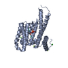
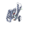
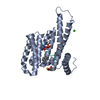
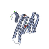
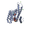
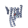
- PDB-3mxn: Crystal structure of the RMI core complex -
+
Open data
ID or keywords:
Loading...
-
Basic information
Entry
Database: PDB / ID: 3mxn
Title
Crystal structure of the RMI core complex
Components
RecQ-mediated genome instability protein 1
RecQ-mediated genome instability protein 2
Keywords
REPLICATION / Bloom syndrome / helicase / RMI / topoisomerase / Replication protein A
Function / homology
Function and homology information
regulation of sister chromatid segregation / reduction of food intake in response to dietary excess / RecQ family helicase-topoisomerase III complex / resolution of DNA recombination intermediates / maintenance of rDNA / resolution of meiotic recombination intermediates / Impaired BRCA2 binding to PALB2 / Defective homologous recombination repair (HRR) due to BRCA1 loss of function / Defective HDR through Homologous Recombination Repair (HRR) due to PALB2 loss of BRCA1 binding function / Defective HDR through Homologous Recombination Repair (HRR) due to PALB2 loss of BRCA2/RAD51/RAD51C binding function ...regulation of sister chromatid segregation / reduction of food intake in response to dietary excess / RecQ family helicase-topoisomerase III complex / resolution of DNA recombination intermediates / maintenance of rDNA / resolution of meiotic recombination intermediates / Impaired BRCA2 binding to PALB2 / Defective homologous recombination repair (HRR) due to BRCA1 loss of function / Defective HDR through Homologous Recombination Repair (HRR) due to PALB2 loss of BRCA1 binding function / Defective HDR through Homologous Recombination Repair (HRR) due to PALB2 loss of BRCA2/RAD51/RAD51C binding function / Homologous DNA Pairing and Strand Exchange / Resolution of D-loop Structures through Synthesis-Dependent Strand Annealing (SDSA) / Resolution of D-loop Structures through Holliday Junction Intermediates / HDR through Single Strand Annealing (SSA) / Impaired BRCA2 binding to RAD51 / Presynaptic phase of homologous DNA pairing and strand exchange / negative regulation of double-strand break repair via homologous recombination / response to glucose / double-strand break repair via homologous recombination / HDR through Homologous Recombination (HRR) / G2/M DNA damage checkpoint / multicellular organism growth / glucose homeostasis / Processing of DNA double-strand break ends / Regulation of TP53 Activity through Phosphorylation / DNA replication / nuclear body / nuclear speck / nucleotide binding / DNA repair / DNA binding / nucleoplasm / nucleus / cytosol Similarity search - Function
Mass: 18.015 Da / Num. of mol.: 251 / Source method: isolated from a natural source / Formula: H2O
-
Experimental details
-
Experiment
Experiment
Method: X-RAY DIFFRACTION / Number of used crystals: 1
-
Sample preparation
Crystal
Density Matthews: 2.06 Å3/Da / Density % sol: 40.23 %
Crystal grow
Temperature: 298 K / Method: vapor diffusion, hanging drop / pH: 7.1 Details: 10 mM Hepes pH 7.1, 0.05 M NaSCN, 20% ethylene glycol, 2 mM DTT, 13% polyethylene glycol 3350, with benzamidine (1%) added to the drop, VAPOR DIFFUSION, HANGING DROP, temperature 298K
Resolution: 1.55→1.58 Å / Redundancy: 5.3 % / Mean I/σ(I) obs: 2.4 / Rsym value: 0.378 / % possible all: 83
-
Processing
Software
Name
Version
Classification
MAR345
datacollection
ARP/wARP
modelbuilding
REFMAC
5.5.0072
refinement
HKL-2000
datareduction
HKL-2000
datascaling
Refinement
Method to determine structure: SAD / Resolution: 1.55→29.8 Å / Cor.coef. Fo:Fc: 0.956 / Cor.coef. Fo:Fc free: 0.938 / SU B: 3.411 / SU ML: 0.057 / Cross valid method: THROUGHOUT / σ(F): 0 / σ(I): 0 / ESU R Free: 0.096 / Stereochemistry target values: MAXIMUM LIKELIHOOD / Details: HYDROGENS HAVE BEEN ADDED IN THE RIDING POSITIONS
Rfactor
Num. reflection
% reflection
Selection details
Rfree
0.23106
1966
5 %
RANDOM
Rwork
0.19548
-
-
-
all
0.1972
40496
-
-
obs
0.1972
37250
96.97 %
-
Solvent computation
Ion probe radii: 0.8 Å / Shrinkage radii: 0.8 Å / VDW probe radii: 1.4 Å / Solvent model: MASK
Displacement parameters
Biso mean: 18.566 Å2
Baniso -1
Baniso -2
Baniso -3
1-
-0.66 Å2
0 Å2
0 Å2
2-
-
0.09 Å2
0 Å2
3-
-
-
0.57 Å2
Refinement step
Cycle: LAST / Resolution: 1.55→29.8 Å
Protein
Nucleic acid
Ligand
Solvent
Total
Num. atoms
2182
0
9
251
2442
Refine LS restraints
Refine-ID
Type
Dev ideal
Dev ideal target
Number
X-RAY DIFFRACTION
r_bond_refined_d
0.008
0.022
2337
X-RAY DIFFRACTION
r_angle_refined_deg
1.191
2.002
3192
X-RAY DIFFRACTION
r_dihedral_angle_1_deg
5.467
5
316
X-RAY DIFFRACTION
r_dihedral_angle_2_deg
32.838
24.149
94
X-RAY DIFFRACTION
r_dihedral_angle_3_deg
13.464
15
436
X-RAY DIFFRACTION
r_dihedral_angle_4_deg
17.879
15
18
X-RAY DIFFRACTION
r_chiral_restr
0.078
0.2
367
X-RAY DIFFRACTION
r_gen_planes_refined
0.005
0.021
1756
X-RAY DIFFRACTION
r_mcbond_it
0.611
1.5
1473
X-RAY DIFFRACTION
r_mcangle_it
1.168
2
2403
X-RAY DIFFRACTION
r_scbond_it
1.877
3
864
X-RAY DIFFRACTION
r_scangle_it
3.181
4.5
771
LS refinement shell
Resolution: 1.55→1.59 Å / Total num. of bins used: 20
Rfactor
Num. reflection
% reflection
Rfree
0.366
130
-
Rwork
0.314
2313
-
obs
-
-
84.3 %
Refinement TLS params.
Method: refined / Refine-ID: X-RAY DIFFRACTION
ID
L11 (°2)
L12 (°2)
L13 (°2)
L22 (°2)
L23 (°2)
L33 (°2)
S11 (Å °)
S12 (Å °)
S13 (Å °)
S21 (Å °)
S22 (Å °)
S23 (Å °)
S31 (Å °)
S32 (Å °)
S33 (Å °)
T11 (Å2)
T12 (Å2)
T13 (Å2)
T22 (Å2)
T23 (Å2)
T33 (Å2)
Origin x (Å)
Origin y (Å)
Origin z (Å)
1
0.3878
0.1068
0.1151
0.1277
0.0383
0.079
-0.0012
-0.0056
-0.0059
0.0079
0.0028
0.0039
-0.0079
0.001
-0.0017
0.0022
0
0.0005
0.0004
0.0003
0.0004
8.1832
0.4551
-10.1492
2
0.4841
-0.023
-0.0482
0.3512
0.074
0.3724
-0.0126
0.0409
-0.0468
-0.0468
0.027
-0.0109
0.0147
0.0239
-0.0144
0.009
-0.0024
0.001
0.009
-0.0074
0.0068
8.2492
-12.8349
-28.479
3
6.2915
-1.3068
-1.3795
9.7117
2.2487
0.7103
-0.1458
0.1272
-0.1986
0.3812
0.0232
0.3984
0.1057
-0.0186
0.1226
0.0167
-0.0003
0.0181
0.011
-0.0017
0.0198
-3.1622
-19.4957
-4.2208
Refinement TLS group
ID
Refine-ID
Refine TLS-ID
Auth asym-ID
Auth seq-ID
1
X-RAY DIFFRACTION
1
A
476 - 625
2
X-RAY DIFFRACTION
2
B
20 - 148
3
X-RAY DIFFRACTION
3
A
1 - 232
+
About Yorodumi
-
News
-
Feb 9, 2022. New format data for meta-information of EMDB entries
New format data for meta-information of EMDB entries
Version 3 of the EMDB header file is now the official format.
The previous official version 1.9 will be removed from the archive.
In the structure databanks used in Yorodumi, some data are registered as the other names, "COVID-19 virus" and "2019-nCoV". Here are the details of the virus and the list of structure data.
Jan 31, 2019. EMDB accession codes are about to change! (news from PDBe EMDB page)
EMDB accession codes are about to change! (news from PDBe EMDB page)
The allocation of 4 digits for EMDB accession codes will soon come to an end. Whilst these codes will remain in use, new EMDB accession codes will include an additional digit and will expand incrementally as the available range of codes is exhausted. The current 4-digit format prefixed with “EMD-” (i.e. EMD-XXXX) will advance to a 5-digit format (i.e. EMD-XXXXX), and so on. It is currently estimated that the 4-digit codes will be depleted around Spring 2019, at which point the 5-digit format will come into force.
The EM Navigator/Yorodumi systems omit the EMD- prefix.
Related info.:Q: What is EMD? / ID/Accession-code notation in Yorodumi/EM Navigator
Yorodumi is a browser for structure data from EMDB, PDB, SASBDB, etc.
This page is also the successor to EM Navigator detail page, and also detail information page/front-end page for Omokage search.
The word "yorodu" (or yorozu) is an old Japanese word meaning "ten thousand". "mi" (miru) is to see.
Related info.:EMDB / PDB / SASBDB / Comparison of 3 databanks / Yorodumi Search / Aug 31, 2016. New EM Navigator & Yorodumi / Yorodumi Papers / Jmol/JSmol / Function and homology information / Changes in new EM Navigator and Yorodumi
 Movie
Movie Controller


 Open data
 Basic information
 Components
 Keywords
 REPLICATION / Bloom syndrome /
  Function and homology information
 Authors
 Citation
 Structure visualization
 Downloads & links
 Other downloads


 PDBj
PDBj


 Assembly


 Mass: 120.152 Da / Num. of mol.: 1 / Source method: obtained synthetically / Formula: C7H8N2
 Mass: 18.015 Da / Num. of mol.: 251 / Source method: isolated from a natural source / Formula: H2O
 Sample preparation
 / Beamline: 21-ID-D / Wavelength: 0.97933, 0.97856
 Processing