Mass: 18.015 Da / Num. of mol.: 107 / Source method: isolated from a natural source / Formula: H2O
Sequence details
THIS CONSTRUCT WAS EXPRESSED WITH A PURIFICATION TAG MGSDKIHHHHHHENLYFQG. THE TAG WAS REMOVED WITH ...THIS CONSTRUCT WAS EXPRESSED WITH A PURIFICATION TAG MGSDKIHHHHHHENLYFQG. THE TAG WAS REMOVED WITH TEV PROTEASE LEAVING ONLY A GLYCINE (0) FOLLOWED BY THE TARGET SEQUENCE.
-
Experimental details
-
Experiment
Experiment
Method: X-RAY DIFFRACTION / Number of used crystals: 1
-
Sample preparation
Crystal
Density Matthews: 2.27 Å3/Da / Density % sol: 45.71 %
Type: MARMOSAIC 325 mm CCD / Detector: CCD / Date: Jun 12, 2009 / Details: Flat mirror (vertical focusing)
Radiation
Monochromator: Single crystal Si(111) bent monochromator (horizontal focusing) Protocol: MAD / Monochromatic (M) / Laue (L): M / Scattering type: x-ray
Radiation wavelength
ID
Wavelength (Å)
Relative weight
1
0.91837
1
2
0.97935
1
3
0.97882
1
Reflection
Resolution: 1.8→29.21 Å / Num. obs: 22224 / % possible obs: 99.9 % / Redundancy: 7.1 % / Biso Wilson estimate: 28 Å2 / Rmerge(I) obs: 0.065 / Rsym value: 0.065 / Net I/σ(I): 18.1
Reflection shell
Resolution (Å)
Redundancy (%)
Rmerge(I) obs
Mean I/σ(I) obs
Num. measured all
Num. unique all
Rsym value
% possible all
1.8-1.85
7.2
0.666
1.2
11519
1610
0.666
100
1.85-1.9
7.2
0.548
1.4
11208
1557
0.548
100
1.9-1.95
7.1
0.417
1.8
10863
1520
0.417
100
1.95-2.01
7.2
0.321
2.4
10712
1496
0.321
100
2.01-2.08
7.2
0.243
3.1
10208
1419
0.243
100
2.08-2.15
7.2
0.187
4.1
10112
1413
0.187
100
2.15-2.23
7.2
0.141
5.3
9669
1351
0.141
100
2.23-2.32
7.1
0.12
6.2
9393
1316
0.12
100
2.32-2.43
7.1
0.103
6.9
8890
1248
0.103
100
2.43-2.55
7.1
0.087
7.7
8464
1184
0.087
100
2.55-2.68
7.1
0.083
8.3
8136
1147
0.083
100
2.68-2.85
7.1
0.078
8.4
7759
1098
0.078
100
2.85-3.04
7.1
0.074
8.4
7205
1018
0.074
100
3.04-3.29
7
0.061
10
6871
977
0.061
100
3.29-3.6
7
0.05
12.3
6170
884
0.05
100
3.6-4.02
7
0.039
15.3
5725
823
0.039
100
4.02-4.65
6.8
0.042
14.3
5010
735
0.042
100
4.65-5.69
6.7
0.045
13.4
4117
618
0.045
100
5.69-8.05
6.3
0.039
15.8
3226
511
0.039
99.9
8.05-29.21
5.4
0.033
18.4
1600
299
0.033
95.7
-
Phasing
Phasing
Method: MAD
-
Processing
Software
Name
Version
Classification
NB
REFMAC
5.6.0059
refinement
PHENIX
refinement
SHELX
phasing
MolProbity
3beta29
modelbuilding
SCALA
3.2.5
datascaling
PDB_EXTRACT
3.006
dataextraction
MOSFLM
datareduction
SHELXD
phasing
autoSHARP
phasing
Refinement
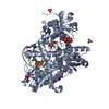
Method to determine structure: MAD / Resolution: 1.8→29.21 Å / Cor.coef. Fo:Fc: 0.967 / Cor.coef. Fo:Fc free: 0.964 / Occupancy max: 1 / Occupancy min: 0.3 / SU B: 4.476 / SU ML: 0.067 / Cross valid method: THROUGHOUT / σ(F): 0 / ESU R Free: 0.1 Stereochemistry target values: MAXIMUM LIKELIHOOD WITH PHASES Details: 1. HYDROGENS HAVE BEEN ADDED IN THE RIDING POSITIONS. 2. ATOM RECORD CONTAINS SUM OF TLS AND RESIDUAL B FACTORS. 3. ANISOU RECORD CONTAINS SUM OF TLS AND RESIDUAL U FACTORS. 4. A MET- ...Details: 1. HYDROGENS HAVE BEEN ADDED IN THE RIDING POSITIONS. 2. ATOM RECORD CONTAINS SUM OF TLS AND RESIDUAL B FACTORS. 3. ANISOU RECORD CONTAINS SUM OF TLS AND RESIDUAL U FACTORS. 4. A MET-INHIBITION PROTOCOL WAS USED FOR SELENOMETHIONINE INCORPORATION DURING PROTEIN EXPRESSION. THE OCCUPANCY OF THE SE ATOMS IN THE MSE RESIDUES WAS REDUCED TO 0.75 FOR THE REDUCED SCATTERING POWER DUE TO PARTIAL S-MET INCORPORATION. 5. CHLORIDE (CL) ION, IMIDAZOLE (IMD), AND ETHYLENE GLYCOL (EDO) MOLECULES FROM THE PURIFICATION/CRYOPROTECTION SOLUTION ARE MODELED. 6. RESIDUES 84-91 HAVE POOR ELECTRON DENSITY SUPPORT.
Rfactor
Num. reflection
% reflection
Selection details
Rfree
0.179
1132
5.1 %
RANDOM
Rwork
0.165
-
-
-
obs
0.165
22179
99.94 %
-
Solvent computation
Ion probe radii: 0.8 Å / Shrinkage radii: 0.8 Å / VDW probe radii: 1.2 Å / Solvent model: BABINET MODEL WITH MASK
In the structure databanks used in Yorodumi, some data are registered as the other names, "COVID-19 virus" and "2019-nCoV". Here are the details of the virus and the list of structure data.
Jan 31, 2019. EMDB accession codes are about to change! (news from PDBe EMDB page)
EMDB accession codes are about to change! (news from PDBe EMDB page)
The allocation of 4 digits for EMDB accession codes will soon come to an end. Whilst these codes will remain in use, new EMDB accession codes will include an additional digit and will expand incrementally as the available range of codes is exhausted. The current 4-digit format prefixed with “EMD-” (i.e. EMD-XXXX) will advance to a 5-digit format (i.e. EMD-XXXXX), and so on. It is currently estimated that the 4-digit codes will be depleted around Spring 2019, at which point the 5-digit format will come into force.
The EM Navigator/Yorodumi systems omit the EMD- prefix.
Related info.:Q: What is EMD? / ID/Accession-code notation in Yorodumi/EM Navigator
Yorodumi is a browser for structure data from EMDB, PDB, SASBDB, etc.
This page is also the successor to EM Navigator detail page, and also detail information page/front-end page for Omokage search.
The word "yorodu" (or yorozu) is an old Japanese word meaning "ten thousand". "mi" (miru) is to see.
Related info.:EMDB / PDB / SASBDB / Comparison of 3 databanks / Yorodumi Search / Aug 31, 2016. New EM Navigator & Yorodumi / Yorodumi Papers / Jmol/JSmol / Function and homology information / Changes in new EM Navigator and Yorodumi
 Movie
Movie Controller

 Yorodumi
 Open data
 Basic information
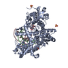
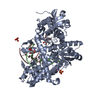
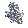
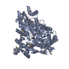
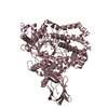
 Components
 Keywords
 transcription regulator / TENA/THI-4/PQQC family /
  Function and homology information
 Ruegeria sp. TM1040 (bacteria)
 Authors
 Citation
 Structure visualization
 Downloads & links
 Other downloads
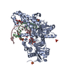

 PDBj
PDBj


 Assembly


 Mass: 69.085 Da / Num. of mol.: 1 / Source method: obtained synthetically / Formula: C3H5N2
 Mass: 62.068 Da / Num. of mol.: 12 / Source method: obtained synthetically / Formula: C2H6O2
 Mass: 35.453 Da / Num. of mol.: 1 / Source method: obtained synthetically / Formula: Cl
 Mass: 18.015 Da / Num. of mol.: 107 / Source method: isolated from a natural source / Formula: H2O
 Sample preparation
 / Beamline: BL11-1 / Wavelength: 0.91837,0.97935,0.97882
 Processing