 Movie
Movie Controller

[English] 日本語
 Yorodumi
- PDB-3mvf: Crystal Structure of Nitrophorin 4 from Rhodnius prolixus Complex... -
+ Open data

- Basic information
| Entry | Database: PDB / ID: 3mvf | ||||||
|---|---|---|---|---|---|---|---|
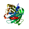
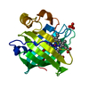
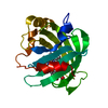
| Title | Crystal Structure of Nitrophorin 4 from Rhodnius prolixus Complexed with Nitrite at pH 7.4 | ||||||
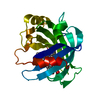
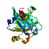
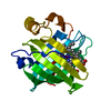
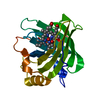
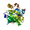
 Components Components | Nitrophorin-4 | ||||||
 Keywords Keywords | TRANSPORT PROTEIN / heme / hemoglobin / iron / lipocalin / nitric oxide / nitrite / nitrophorin | ||||||
| Function / homology |  Function and homology informationnitrite dismutase / histamine binding / nitric oxide binding / vasodilation / oxidoreductase activity / extracellular region / metal ion binding Function and homology informationnitrite dismutase / histamine binding / nitric oxide binding / vasodilation / oxidoreductase activity / extracellular region / metal ion bindingSimilarity search - Function | ||||||
| Biological species |   Rhodnius prolixus (insect) Rhodnius prolixus (insect) | ||||||
| Method | X-RAY DIFFRACTION / SYNCHROTRON / MOLECULAR REPLACEMENT / Resolution: 1.4 Å | ||||||
 Authors Authors | Ogata, H. / He, C. / Knipp, M. | ||||||
 Citation Citation | Journal: Biochemistry / Year: 2010 Title: Formation of the complex of nitrite with the ferriheme b beta-barrel proteins nitrophorin 4 and nitrophorin 7. Authors: He, C. / Ogata, H. / Knipp, M. | ||||||
| History |
|
- Structure visualization
| Structure viewer | Molecule: MolmilJmol/JSmol |
|---|
- Downloads & links
-Download
| PDBx/mmCIF format | 3mvf.cif.gz | 92.3 KB | Display | PDBx/mmCIF format |
|---|---|---|---|---|
| PDB format | pdb3mvf.ent.gz | 75.6 KB | Display | PDB format |
| PDBx/mmJSON format | 3mvf.json.gz | Tree view | PDBx/mmJSON format | |
| Others |  Other downloads Other downloads |
-Validation report
| Arichive directory | https://data.pdbj.org/pub/pdb/validation_reports/mv/3mvfftp://data.pdbj.org/pub/pdb/validation_reports/mv/3mvf | HTTPS FTP |
|---|
-Related structure data
| Similar structure data |
|---|
-Links
 PDBj
PDBj


- Assembly
| Deposited unit | 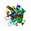
| ||||||||
|---|---|---|---|---|---|---|---|---|---|
| 1 |
| ||||||||
| 2 | 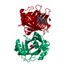
| ||||||||
| Unit cell |
| ||||||||
| Components on special symmetry positions |
|
-Components
| #1: Protein | / NP4 Mass: 20292.664 Da / Num. of mol.: 1 Source method: isolated from a genetically manipulated source Source: (gene. exp.) Rhodnius prolixus (insect) / Plasmid: pET17 / Production host:  Escherichia coli (E. coli) / Strain (production host): BL21 (DE3) / References: UniProt: Q94734 Escherichia coli (E. coli) / Strain (production host): BL21 (DE3) / References: UniProt: Q94734 | ||
|---|---|---|---|
| #2: Chemical | ChemComp-HEM / Heme B  Mass: 616.487 Da / Num. of mol.: 1 / Source method: obtained synthetically / Formula: C34H32FeN4O4 Mass: 616.487 Da / Num. of mol.: 1 / Source method: obtained synthetically / Formula: C34H32FeN4O4 | ||
| #3: Chemical | Nitrite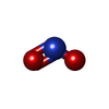  Mass: 46.005 Da / Num. of mol.: 2 / Source method: obtained synthetically / Formula: NO2 Mass: 46.005 Da / Num. of mol.: 2 / Source method: obtained synthetically / Formula: NO2#4: Water | ChemComp-HOH / | Water Mass: 18.015 Da / Num. of mol.: 223 / Source method: isolated from a natural source / Formula: H2O Mass: 18.015 Da / Num. of mol.: 223 / Source method: isolated from a natural source / Formula: H2O |
-Experimental details
-Experiment
| Experiment | Method: X-RAY DIFFRACTION / Number of used crystals: 1 |
|---|
- Sample preparation
| Crystal | Density Matthews: 1.94 Å3/Da / Density % sol: 36.75 % |
|---|---|
| Crystal grow | Temperature: 297 K / Method: vapor diffusion, sitting drop / pH: 7.4 Details: Ammonium phosphate, pH 7.4, VAPOR DIFFUSION, SITTING DROP, temperature 297K |
-Data collection
| Diffraction | Mean temperature: 100 K |
|---|---|
| Diffraction source | Source: SYNCHROTRON / Site: BESSY  / Beamline: 14.2 / Wavelength: 0.91892 Å / Beamline: 14.2 / Wavelength: 0.91892 Å |
| Detector | Type: MARMOSAIC 225 mm CCD / Detector: CCD / Date: Mar 23, 2010 |
| Radiation | Monochromator: Si / Protocol: SINGLE WAVELENGTH / Monochromatic (M) / Laue (L): M / Scattering type: x-ray |
| Radiation wavelength | Wavelength: 0.91892 Å / Relative weight: 1 |
| Reflection | Resolution: 1.4→30.53 Å / Num. obs: 30694 / % possible obs: 97.7 % / Observed criterion σ(F): 0 / Observed criterion σ(I): 0 / Rmerge(I) obs: 0.038 |
| Reflection shell | Resolution: 1.4→1.44 Å / Rmerge(I) obs: 0.193 / % possible all: 93.7 |
- Processing
| Software |
| ||||||||||||||||||||||||||||||||||||||||||||||||||||||||||||||||||||||
|---|---|---|---|---|---|---|---|---|---|---|---|---|---|---|---|---|---|---|---|---|---|---|---|---|---|---|---|---|---|---|---|---|---|---|---|---|---|---|---|---|---|---|---|---|---|---|---|---|---|---|---|---|---|---|---|---|---|---|---|---|---|---|---|---|---|---|---|---|---|---|---|
| Refinement | Method to determine structure: MOLECULAR REPLACEMENT / Resolution: 1.4→15 Å / Cor.coef. Fo:Fc: 0.973 / Cor.coef. Fo:Fc free: 0.954 / SU B: 1.765 / SU ML: 0.033 / Cross valid method: THROUGHOUT / σ(F): 0 / ESU R Free: 0.064 / Stereochemistry target values: MAXIMUM LIKELIHOOD
| ||||||||||||||||||||||||||||||||||||||||||||||||||||||||||||||||||||||
| Solvent computation | Ion probe radii: 0.8 Å / Shrinkage radii: 0.8 Å / VDW probe radii: 1.4 Å / Solvent model: MASK | ||||||||||||||||||||||||||||||||||||||||||||||||||||||||||||||||||||||
| Displacement parameters | Biso mean: 16.999 Å2
| ||||||||||||||||||||||||||||||||||||||||||||||||||||||||||||||||||||||
| Refinement step | Cycle: LAST / Resolution: 1.4→15 Å
| ||||||||||||||||||||||||||||||||||||||||||||||||||||||||||||||||||||||
| Refine LS restraints |
| ||||||||||||||||||||||||||||||||||||||||||||||||||||||||||||||||||||||
| LS refinement shell | Resolution: 1.4→1.436 Å / Total num. of bins used: 20
| ||||||||||||||||||||||||||||||||||||||||||||||||||||||||||||||||||||||
| Refinement TLS params. | Method: refined / Origin x: -8.2211 Å / Origin y: -0.3259 Å / Origin z: -13.2321 Å
|
