 Movie
Movie Controller

[English] 日本語
 Yorodumi
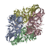
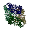
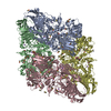
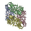
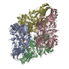
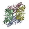
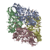
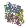
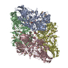
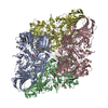
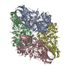
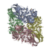
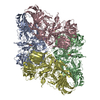
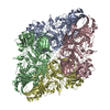
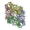
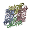
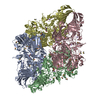
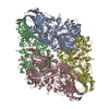
- PDB-3mv1: E.Coli (lacZ) beta-galactosidase (R599A) in complex with Guanidinium -
+ Open data

- Basic information
| Entry | Database: PDB / ID: 3mv1 | ||||||
|---|---|---|---|---|---|---|---|
| Title | E.Coli (lacZ) beta-galactosidase (R599A) in complex with Guanidinium | ||||||
 Components Components | Beta-galactosidase | ||||||
 Keywords Keywords | HYDROLASE / Arg-599-Ala / BETA-GALACTOSIDASE / HYDROLASE TIM BARREL(ALPHA/BETA BARREL) / JELLY-ROLL BARREL / IMMUNOGLOBULIN BETA SUPERSANDWHICH / GLYCOSIDASE | ||||||
| Function / homology |  Function and homology information Function and homology informationalkali metal ion binding / organic substance catabolic process / lactose catabolic process / beta-galactosidase complex / beta-galactosidase / beta-galactosidase activity / carbohydrate binding / carbohydrate metabolic process / magnesium ion binding / identical protein bindingSimilarity search - Function | ||||||
| Biological species |  Escherichia coli (E. coli) Escherichia coli (E. coli) | ||||||
| Method | X-RAY DIFFRACTION / SYNCHROTRON / MOLECULAR REPLACEMENT / Resolution: 2.2 Å | ||||||
 Authors Authors | Dugdale, M.L. / Vance, M. / Driedger, M.L. / Nibber, A. / Tran, A. / Huber, R.E. | ||||||
 Citation Citation | Journal: Biochem.Cell Biol. / Year: 2010 Title: Importance of Arg-599 of b-galactosidase (Escherichia coli) as an anchor for the open conformations of Phe-601 and the active-site loop Authors: Dugdale, M.L. / Vance, M.L. / Wheatley, R.W. / Driedger, M.R. / Nibber, A. / Tran, A. / Huber, R.E. | ||||||
| History |
|
- Structure visualization
| Structure viewer | Molecule: MolmilJmol/JSmol |
|---|
- Downloads & links
-Download
| PDBx/mmCIF format | 3mv1.cif.gz | 922.1 KB | Display | PDBx/mmCIF format |
|---|---|---|---|---|
| PDB format | pdb3mv1.ent.gz | 736.7 KB | Display | PDB format |
| PDBx/mmJSON format | 3mv1.json.gz | Tree view | PDBx/mmJSON format | |
| Others |  Other downloads Other downloads |
-Validation report
| Arichive directory | https://data.pdbj.org/pub/pdb/validation_reports/mv/3mv1ftp://data.pdbj.org/pub/pdb/validation_reports/mv/3mv1 | HTTPS FTP |
|---|
-Related structure data
| Related structure data |  3muzC  3mv0C 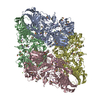 1dp0S S: Starting model for refinement C: citing same article ( |
|---|---|
| Similar structure data |


-Links
 PDBj
PDBj


- Assembly
| Deposited unit | 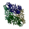
| ||||||||
|---|---|---|---|---|---|---|---|---|---|
| 1 |
| ||||||||
| Unit cell |
|
-Components
-Protein , 1 types, 4 molecules 1234
| #1: Protein | Mass: 119657.633 Da / Num. of mol.: 4 / Mutation: R599A Source method: isolated from a genetically manipulated source Source: (gene. exp.) Escherichia coli (E. coli) / Strain: K12 / Gene: lacZ / Plasmid: pBAD/HIS/LacZ / Production host: Escherichia coli (E. coli) / Strain (production host): LMG194References: UniProt: B8LFD6, UniProt: P00722*PLUS, beta-galactosidase |
|---|
-Non-polymers , 5 types, 3795 molecules 

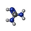


| #2: Chemical | ChemComp-MG /  Mass: 24.305 Da / Num. of mol.: 12 / Source method: obtained synthetically / Formula: Mg Mass: 24.305 Da / Num. of mol.: 12 / Source method: obtained synthetically / Formula: Mg#3: Chemical | ChemComp-NA /  Mass: 22.990 Da / Num. of mol.: 16 / Source method: obtained synthetically / Formula: Na Mass: 22.990 Da / Num. of mol.: 16 / Source method: obtained synthetically / Formula: Na#4: Chemical | ChemComp-GAI / Guanidine Mass: 59.070 Da / Num. of mol.: 4 / Source method: obtained synthetically / Formula: CH5N3 Mass: 59.070 Da / Num. of mol.: 4 / Source method: obtained synthetically / Formula: CH5N3#5: Chemical | ChemComp-DMS / Dimethyl sulfoxide Mass: 78.133 Da / Num. of mol.: 109 / Source method: obtained synthetically / Formula: C2H6OS / Comment: DMSO, precipitant*YM Mass: 78.133 Da / Num. of mol.: 109 / Source method: obtained synthetically / Formula: C2H6OS / Comment: DMSO, precipitant*YM#6: Water | ChemComp-HOH / | WaterMass: 18.015 Da / Num. of mol.: 3654 / Source method: isolated from a natural source / Formula: H2O |
|---|
-Experimental details
-Experiment
| Experiment | Method: X-RAY DIFFRACTION / Number of used crystals: 1 |
|---|
- Sample preparation
| Crystal | Density Matthews: 2.61 Å3/Da / Density % sol: 52.84 % |
|---|---|
| Crystal grow | Temperature: 288 K / Method: vapor diffusion, hanging drop / pH: 6.5 Details: PEG8000, NaCl, MgCl2, DTT, Bis-Tris, pH6.5, VAPOR DIFFUSION, HANGING DROP, temperature 288K |
-Data collection
| Diffraction | Mean temperature: 100 K |
|---|---|
| Diffraction source | Source: SYNCHROTRON / Site: ALS  / Beamline: 8.3.1 / Wavelength: 1.11587 Å / Beamline: 8.3.1 / Wavelength: 1.11587 Å |
| Detector | Type: ADSC QUANTUM 210 / Detector: CCD / Date: Jul 17, 2008 / Details: KOHZU: Double Crystal SI(111) |
| Radiation | Monochromator: SI(111) / Protocol: SINGLE WAVELENGTH / Monochromatic (M) / Laue (L): M / Scattering type: x-ray |
| Radiation wavelength | Wavelength: 1.11587 Å / Relative weight: 1 |
| Reflection | Resolution: 2.2→71.29 Å / Num. all: 251514 / Num. obs: 251768 / % possible obs: 99.7 % / Observed criterion σ(F): 0 / Observed criterion σ(I): 0 / Redundancy: 3.06 % / Biso Wilson estimate: 19 Å2 / Limit h max: 68 / Limit h min: 0 / Limit k max: 73 / Limit k min: 0 / Limit l max: 92 / Limit l min: 0 / Observed criterion F max: 5196848.8 / Observed criterion F min: 19.171 / Rmerge(I) obs: 0.104 / Net I/σ(I): 7.7 |
| Reflection shell | Resolution: 2.2→2.32 Å / Redundancy: 3.06 % / Rmerge(I) obs: 0.47 / Mean I/σ(I) obs: 3 / Num. unique all: 36156 / % possible all: 98.9 |
- Processing
| Software |
| ||||||||||||||||||||||||||||||||||||||||||||||||||||||||||||||||||||||||||||||||||||||||||
|---|---|---|---|---|---|---|---|---|---|---|---|---|---|---|---|---|---|---|---|---|---|---|---|---|---|---|---|---|---|---|---|---|---|---|---|---|---|---|---|---|---|---|---|---|---|---|---|---|---|---|---|---|---|---|---|---|---|---|---|---|---|---|---|---|---|---|---|---|---|---|---|---|---|---|---|---|---|---|---|---|---|---|---|---|---|---|---|---|---|---|---|
| Refinement | Method to determine structure: MOLECULAR REPLACEMENT Starting model: PDB entry 1DP0 Resolution: 2.2→71.29 Å / Rfactor Rfree error: 0.004 / Occupancy max: 1 / Occupancy min: 1 / Isotropic thermal model: Isotropic / Cross valid method: THROUGHOUT / σ(F): 0 / σ(I): 0 / Stereochemistry target values: Engh & Huber
| ||||||||||||||||||||||||||||||||||||||||||||||||||||||||||||||||||||||||||||||||||||||||||
| Solvent computation | Solvent model: CNS bulk solvent model used / Bsol: 86.7379 Å2 / ksol: 0.4 e/Å3 | ||||||||||||||||||||||||||||||||||||||||||||||||||||||||||||||||||||||||||||||||||||||||||
| Displacement parameters | Biso max: 109.12 Å2 / Biso mean: 27.14 Å2 / Biso min: 2.23 Å2
| ||||||||||||||||||||||||||||||||||||||||||||||||||||||||||||||||||||||||||||||||||||||||||
| Refine analyze |
| ||||||||||||||||||||||||||||||||||||||||||||||||||||||||||||||||||||||||||||||||||||||||||
| Refinement step | Cycle: LAST / Resolution: 2.2→71.29 Å
| ||||||||||||||||||||||||||||||||||||||||||||||||||||||||||||||||||||||||||||||||||||||||||
| Refine LS restraints |
| ||||||||||||||||||||||||||||||||||||||||||||||||||||||||||||||||||||||||||||||||||||||||||
| LS refinement shell | Refine-ID: X-RAY DIFFRACTION
|
