 Movie
Movie Controller


+ Open data

- Basic information
| Entry | Database: PDB / ID: 3ms6 | ||||||
|---|---|---|---|---|---|---|---|
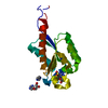
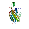
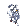
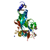
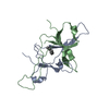
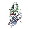
| Title | Crystal structure of Hepatitis B X-Interacting Protein (HBXIP) | ||||||
 Components Components | Hepatitis B virus X-interacting protein | ||||||
 Keywords Keywords |  PROTEIN BINDING / alpha-beta protein / profilin-like fold / Roadblock/LC7 domain superfamily PROTEIN BINDING / alpha-beta protein / profilin-like fold / Roadblock/LC7 domain superfamily | ||||||
| Function / homology |  Function and homology information Function and homology informationpositive regulation of RNA polymerase II regulatory region sequence-specific DNA binding / FNIP-folliculin RagC/D GAP / Ragulator complex / protein localization to lysosome / TORC1 signaling / MTOR signalling / Amino acids regulate mTORC1 / Energy dependent regulation of mTOR by LKB1-AMPK / Macroautophagy / regulation of cell size ...positive regulation of RNA polymerase II regulatory region sequence-specific DNA binding / FNIP-folliculin RagC/D GAP / Ragulator complex / protein localization to lysosome / TORC1 signaling / MTOR signalling / Amino acids regulate mTORC1 / Energy dependent regulation of mTOR by LKB1-AMPK / Macroautophagy / regulation of cell size / mTORC1-mediated signalling / positive regulation of TOR signaling / positive regulation of TORC1 signaling / viral genome replication / Regulation of PTEN gene transcription / positive regulation of interleukin-8 production / cellular response to amino acid stimulus / TP53 Regulates Metabolic Genes / response to virus / negative regulation of cysteine-type endopeptidase activity involved in apoptotic process / positive regulation of protein localization to nucleus / late endosome membrane / positive regulation of NF-kappaB transcription factor activity / positive regulation of canonical NF-kappaB signal transduction / lysosome / lysosomal membrane / positive regulation of gene expression / protein-containing complex / cytosolSimilarity search - Function | ||||||
| Biological species |  Homo sapiens (human) Homo sapiens (human) | ||||||
| Method | X-RAY DIFFRACTION / SYNCHROTRON / SAD / Resolution: 2.085 Å | ||||||
 Authors Authors | Garcia-Saez, I. / Skoufias, D. | ||||||
 Citation Citation | Journal: J.Mol.Biol. / Year: 2011 Title: Structural Characterization of HBXIP: The Protein That Interacts with the Anti-Apoptotic Protein Survivin and the Oncogenic Viral Protein HBx. Authors: Garcia-Saez, I. / Lacroix, F.B. / Blot, D. / Gabel, F. / Skoufias, D.A. | ||||||
| History |
|
- Structure visualization
| Structure viewer | Molecule: MolmilJmol/JSmol |
|---|
- Downloads & links
-Download
| PDBx/mmCIF format | 3ms6.cif.gz | 27.2 KB | Display | PDBx/mmCIF format |
|---|---|---|---|---|
| PDB format | pdb3ms6.ent.gz | 20.5 KB | Display | PDB format |
| PDBx/mmJSON format | 3ms6.json.gz | Tree view | PDBx/mmJSON format | |
| Others |  Other downloads Other downloads |
-Validation report
| Arichive directory | https://data.pdbj.org/pub/pdb/validation_reports/ms/3ms6ftp://data.pdbj.org/pub/pdb/validation_reports/ms/3ms6 | HTTPS FTP |
|---|
-Related structure data


-Links
 PDBj
PDBj


- Assembly
| Deposited unit | 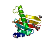
| ||||||||
|---|---|---|---|---|---|---|---|---|---|
| 1 | 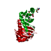
| ||||||||
| Unit cell |
|
-Components
| #1: Protein | Mass: 10881.634 Da / Num. of mol.: 1 Source method: isolated from a genetically manipulated source Source: (gene. exp.) Homo sapiens (human) / Gene: HBXIP, XIP / Plasmid: pET-23d(+) / Production host:  Escherichia coli (E. coli) / Strain (production host): BL21(DE3) / References: UniProt: O43504 Escherichia coli (E. coli) / Strain (production host): BL21(DE3) / References: UniProt: O43504 | ||||
|---|---|---|---|---|---|
| #2: Chemical | ChemComp-PEG / Diethylene glycol  Mass: 106.120 Da / Num. of mol.: 1 / Source method: obtained synthetically / Formula: C4H10O3 Mass: 106.120 Da / Num. of mol.: 1 / Source method: obtained synthetically / Formula: C4H10O3 | ||||
| #3: Chemical | Isopropyl alcohol  Mass: 60.095 Da / Num. of mol.: 2 / Source method: obtained synthetically / Formula: C3H8O / Comment: alkaloid*YM Mass: 60.095 Da / Num. of mol.: 2 / Source method: obtained synthetically / Formula: C3H8O / Comment: alkaloid*YM#4: Chemical | ChemComp-GOL / | Glycerol  Mass: 92.094 Da / Num. of mol.: 1 / Source method: obtained synthetically / Formula: C3H8O3 Mass: 92.094 Da / Num. of mol.: 1 / Source method: obtained synthetically / Formula: C3H8O3#5: Water | ChemComp-HOH / | Water Mass: 18.015 Da / Num. of mol.: 42 / Source method: isolated from a natural source / Formula: H2O Mass: 18.015 Da / Num. of mol.: 42 / Source method: isolated from a natural source / Formula: H2O |
-Experimental details
-Experiment
| Experiment | Method: X-RAY DIFFRACTION / Number of used crystals: 1 |
|---|
- Sample preparation
| Crystal | Density Matthews: 1.98 Å3/Da / Density % sol: 37.75 % |
|---|---|
| Crystal grow | Temperature: 292 K / Method: vapor diffusion, hanging drop / pH: 5.6 Details: 20-24% PEG3350, 20% isopropanol, 0.1M tri-sodium citrate dihydrate, pH 5.6, VAPOR DIFFUSION, HANGING DROP, temperature 292K |
-Data collection
| Diffraction | Mean temperature: 100 K |
|---|---|
| Diffraction source | Source: SYNCHROTRON / Site: ESRF  / Beamline: ID14-4 / Wavelength: 0.979 Å / Beamline: ID14-4 / Wavelength: 0.979 Å |
| Detector | Type: ADSC QUANTUM 315r / Detector: CCD / Date: Apr 30, 2009 / Details: Toroidal focusing mirror |
| Radiation | Monochromator: Single Silicon (111) monochromator / Protocol: MAD / Monochromatic (M) / Laue (L): M / Scattering type: x-ray |
| Radiation wavelength | Wavelength: 0.979 Å / Relative weight: 1 |
| Reflection | Resolution: 2.085→48.997 Å / Num. all: 5623 / Num. obs: 5586 / % possible obs: 99.6 % / Observed criterion σ(I): 1 / Redundancy: 12.9 % / Biso Wilson estimate: 25 Å2 / Rmerge(I) obs: 0.096 / Rsym value: 0.093 / Net I/σ(I): 6.7 |
| Reflection shell | Resolution: 2.085→2.2 Å / Redundancy: 9.7 % / Rmerge(I) obs: 0.4 / Mean I/σ(I) obs: 5.9 / Num. unique all: 774 / Rsym value: 0.38 / % possible all: 97.5 |
- Processing
| Software |
| ||||||||||||||||||||||||
|---|---|---|---|---|---|---|---|---|---|---|---|---|---|---|---|---|---|---|---|---|---|---|---|---|---|
| Refinement | Method to determine structure: SAD / Resolution: 2.085→28.918 Å / SU ML: 0.29 / σ(F): 1.38 / Stereochemistry target values: ML
| ||||||||||||||||||||||||
| Solvent computation | Shrinkage radii: 0.9 Å / VDW probe radii: 1.11 Å / Solvent model: FLAT BULK SOLVENT MODEL / Bsol: 68.09 Å2 / ksol: 0.413 e/Å3 | ||||||||||||||||||||||||
| Displacement parameters |
| ||||||||||||||||||||||||
| Refinement step | Cycle: LAST / Resolution: 2.085→28.918 Å
| ||||||||||||||||||||||||
| Refine LS restraints |
| ||||||||||||||||||||||||
| LS refinement shell |
|
