 Movie
Movie Controller

[English] 日本語
 Yorodumi
- PDB-3mre: Crystal Structure of MHC class I HLA-A2 molecule complexed with E... -
+ Open data

- Basic information
| Entry | Database: PDB / ID: 3mre | ||||||
|---|---|---|---|---|---|---|---|
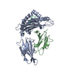
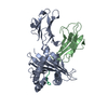
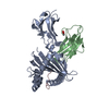
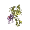
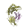
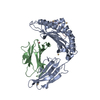
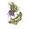
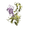
| Title | Crystal Structure of MHC class I HLA-A2 molecule complexed with EBV bmlf1-280-288 nonapeptide | ||||||
 Components Components |
| ||||||
 Keywords Keywords |  IMMUNE SYSTEM / MHC class I / HLA / IMMUNE RESPONSE / NONAPEPTIDE / VIRAL PEPTIDE / EPSTEIN-BARR VIRUS / BMLF1 PROTEIN / EB2 protein IMMUNE SYSTEM / MHC class I / HLA / IMMUNE RESPONSE / NONAPEPTIDE / VIRAL PEPTIDE / EPSTEIN-BARR VIRUS / BMLF1 PROTEIN / EB2 protein | ||||||
| Function / homology |  Function and homology information Function and homology informationsymbiont-mediated suppression of host PKR/eIFalpha signaling / protein serine/threonine kinase inhibitor activity / T cell mediated cytotoxicity directed against tumor cell target / antigen processing and presentation of endogenous peptide antigen via MHC class I via ER pathway, TAP-dependent / positive regulation of memory T cell activation / TAP complex binding / antigen processing and presentation of exogenous peptide antigen via MHC class I / Golgi medial cisterna / positive regulation of CD8-positive, alpha-beta T cell activation / CD8-positive, alpha-beta T cell activation ...symbiont-mediated suppression of host PKR/eIFalpha signaling / protein serine/threonine kinase inhibitor activity / T cell mediated cytotoxicity directed against tumor cell target / antigen processing and presentation of endogenous peptide antigen via MHC class I via ER pathway, TAP-dependent / positive regulation of memory T cell activation / TAP complex binding / antigen processing and presentation of exogenous peptide antigen via MHC class I / Golgi medial cisterna / positive regulation of CD8-positive, alpha-beta T cell activation / CD8-positive, alpha-beta T cell activation / positive regulation of CD8-positive, alpha-beta T cell proliferation / CD8 receptor binding / mRNA transport / endoplasmic reticulum exit site / beta-2-microglobulin binding / TAP binding / protection from natural killer cell mediated cytotoxicity / antigen processing and presentation of endogenous peptide antigen via MHC class I via ER pathway, TAP-independent / antigen processing and presentation of endogenous peptide antigen via MHC class Ib / detection of bacterium / T cell receptor binding / positive regulation of ferrous iron binding / positive regulation of transferrin receptor binding / positive regulation of receptor binding / early endosome lumen / Nef mediated downregulation of MHC class I complex cell surface expression / DAP12 interactions / negative regulation of receptor binding / lumenal side of endoplasmic reticulum membrane / Endosomal/Vacuolar pathway / Antigen Presentation: Folding, assembly and peptide loading of class I MHC / antigen processing and presentation of exogenous protein antigen via MHC class Ib, TAP-dependent / cellular response to iron(III) ion / negative regulation of forebrain neuron differentiation / ER to Golgi transport vesicle membrane / peptide antigen assembly with MHC class I protein complex / response to molecule of bacterial origin / regulation of erythrocyte differentiation / regulation of iron ion transport / MHC class I peptide loading complex / HFE-transferrin receptor complex / T cell mediated cytotoxicity / cellular response to iron ion / antigen processing and presentation of endogenous peptide antigen via MHC class I / positive regulation of T cell cytokine production / MHC class I protein complex / multicellular organismal-level iron ion homeostasis / positive regulation of T cell mediated cytotoxicity / peptide antigen assembly with MHC class II protein complex / negative regulation of neurogenesis / MHC class II protein complex / positive regulation of receptor-mediated endocytosis / cellular response to nicotine / recycling endosome membrane / specific granule lumen / phagocytic vesicle membrane / peptide antigen binding / positive regulation of cellular senescence / antigen processing and presentation of exogenous peptide antigen via MHC class II / Immunoregulatory interactions between a Lymphoid and a non-Lymphoid cell / positive regulation of immune response / negative regulation of epithelial cell proliferation / Interferon gamma signaling / positive regulation of T cell activation / Modulation by Mtb of host immune system / Interferon alpha/beta signaling / positive regulation of type II interferon production / sensory perception of smell / E3 ubiquitin ligases ubiquitinate target proteins / negative regulation of neuron projection development / tertiary granule lumen / DAP12 signaling / MHC class II protein complex binding / T cell differentiation in thymus / late endosome membrane / positive regulation of protein binding / ER-Phagosome pathway / antibacterial humoral response / iron ion transport / T cell receptor signaling pathway / protein refolding / early endosome membrane / protein homotetramerization / intracellular iron ion homeostasis / host cell cytoplasm / amyloid fibril formation / learning or memory / defense response to Gram-positive bacterium / immune response / Amyloid fiber formation / lysosomal membrane / external side of plasma membrane / endoplasmic reticulum lumen / Golgi membrane / signaling receptor binding / focal adhesion / innate immune response / symbiont-mediated suppression of host type I interferon-mediated signaling pathway / host cell nucleus / Neutrophil degranulationSimilarity search - Function | ||||||
| Biological species |  Homo sapiens (human) Homo sapiens (human) Epstein-Barr virus EBV (Epstein-Barr virus) Epstein-Barr virus EBV (Epstein-Barr virus) | ||||||
| Method | X-RAY DIFFRACTION / SYNCHROTRON / MOLECULAR REPLACEMENT / Resolution: 1.1 Å | ||||||
 Authors Authors | Gras, S. / Echasserieau, K. / Saulquin, X. / Bonneville, M. / Housset, D. | ||||||
 Citation Citation | Journal: J.Immunol. / Year: 2014 Title: Analysis of Relationships between Peptide/MHC Structural Features and Naive T Cell Frequency in Humans. Authors: Reiser, J.B. / Legoux, F. / Gras, S. / Trudel, E. / Chouquet, A. / Leger, A. / Le Gorrec, M. / Machillot, P. / Bonneville, M. / Saulquin, X. / Housset, D. | ||||||
| History |
|
- Structure visualization
| Structure viewer | Molecule: MolmilJmol/JSmol |
|---|
- Downloads & links
-Download
| PDBx/mmCIF format | 3mre.cif.gz | 121 KB | Display | PDBx/mmCIF format |
|---|---|---|---|---|
| PDB format | pdb3mre.ent.gz | 92.3 KB | Display | PDB format |
| PDBx/mmJSON format | 3mre.json.gz | Tree view | PDBx/mmJSON format | |
| Others |  Other downloads Other downloads |
-Validation report
| Arichive directory | https://data.pdbj.org/pub/pdb/validation_reports/mr/3mreftp://data.pdbj.org/pub/pdb/validation_reports/mr/3mre | HTTPS FTP |
|---|
-Related structure data
| Related structure data |  3mrcC  3mrdC  3mrgC  3mrhC  3mrlC  3mroC  3mrrC  3gsoS C: citing same article ( S: Starting model for refinement |
|---|---|
| Similar structure data |


-Links
 PDBj
PDBj

- Assembly
| Deposited unit | 
| ||||||||
|---|---|---|---|---|---|---|---|---|---|
| 1 |
| ||||||||
| Unit cell |
|
-Components
| #1: Protein | Mass: 34008.711 Da / Num. of mol.: 1 / Fragment: HLA-A*0201 alpha chain, UNP resiude 25-300 / Mutation: A245V Source method: isolated from a genetically manipulated source Details: C-terminal biotin acceptor peptide sequence tag (GSLHHILDAQKMVWNHR) Source: (gene. exp.) Homo sapiens (human) / Gene: HLA, HLA-A, HLAA / Plasmid: pHN1 / Production host:  Escherichia coli (E. coli) / Strain (production host): X90F LAQQ1 / References: UniProt: P01892 Escherichia coli (E. coli) / Strain (production host): X90F LAQQ1 / References: UniProt: P01892 |
|---|---|
| #2: Protein | Beta-2 microglobulin / Beta-2-microglobulin form pI 5.3 Mass: 11879.356 Da / Num. of mol.: 1 Source method: isolated from a genetically manipulated source Source: (gene. exp.) Homo sapiens (human) / Gene: B2M, BETA-2 MICROGLUBULIN, CDABP0092, HDCMA22P / Plasmid: pHN1 / Production host: Escherichia coli (E. coli) / Strain (production host): X90F LAQQ1 / References: UniProt: P61769 |
| #3: Protein/peptide | Mass: 920.191 Da / Num. of mol.: 1 Fragment: EB2 PROTEIN FRAGMENT (BMLF1 280-288), UNP residues 300-308 Source method: obtained synthetically / Details: chemical synthesis Source: (synth.) Epstein-Barr virus EBV (Epstein-Barr virus)References: UniProt: Q3KSU1 |
| #4: Water | ChemComp-HOH / Water Mass: 18.015 Da / Num. of mol.: 685 / Source method: isolated from a natural source / Formula: H2O Mass: 18.015 Da / Num. of mol.: 685 / Source method: isolated from a natural source / Formula: H2O |
| Compound details | EBV BSFL2 AND BMLF1 ORFS ARE JOINED TO ENCODE THE EB2 PROTEIN. EB2 PROTEIN IS ALSO CALLED ...EBV BSFL2 AND BMLF1 ORFS ARE JOINED TO ENCODE THE EB2 PROTEIN. EB2 PROTEIN IS ALSO CALLED BSFL2/BMLF1, MTA OR SM. |
| Sequence details | THE GLCTLVAML NONAPEPTIDE WAS FIRST IDENTIFIED AS AN HLA-A2 RESTRICTED T CELL EPITOPE ON A ...THE GLCTLVAML NONAPEPTID |
-Experimental details
-Experiment
| Experiment | Method: X-RAY DIFFRACTION / Number of used crystals: 1 |
|---|
- Sample preparation
| Crystal | Density Matthews: 2.25 Å3/Da / Density % sol: 45.32 % |
|---|---|
| Crystal grow | Temperature: 293 K / Method: vapor diffusion, hanging drop / pH: 6.5 Details: 15% PEG 6000, 0.1M NaCitrate, 0.1M NaCl, 5mg/ml protein conc., pH 6.5, VAPOR DIFFUSION, HANGING DROP, temperature 293K |
-Data collection
| Diffraction | Mean temperature: 100 K | |||||||||||||||||||||||||||||||||||||||||||||||||||||||||||||||||||||||||||||||||||||||||||||||||||||||||||||||||||||||||||||||||||||||||||||||||||
|---|---|---|---|---|---|---|---|---|---|---|---|---|---|---|---|---|---|---|---|---|---|---|---|---|---|---|---|---|---|---|---|---|---|---|---|---|---|---|---|---|---|---|---|---|---|---|---|---|---|---|---|---|---|---|---|---|---|---|---|---|---|---|---|---|---|---|---|---|---|---|---|---|---|---|---|---|---|---|---|---|---|---|---|---|---|---|---|---|---|---|---|---|---|---|---|---|---|---|---|---|---|---|---|---|---|---|---|---|---|---|---|---|---|---|---|---|---|---|---|---|---|---|---|---|---|---|---|---|---|---|---|---|---|---|---|---|---|---|---|---|---|---|---|---|---|---|---|---|
| Diffraction source | Source: SYNCHROTRON / Site: ESRF  / Beamline: ID14-3 / Wavelength: 0.931 Å / Beamline: ID14-3 / Wavelength: 0.931 Å | |||||||||||||||||||||||||||||||||||||||||||||||||||||||||||||||||||||||||||||||||||||||||||||||||||||||||||||||||||||||||||||||||||||||||||||||||||
| Detector | Type: MAR CCD 165 mm / Detector: CCD / Date: Dec 16, 2005 | |||||||||||||||||||||||||||||||||||||||||||||||||||||||||||||||||||||||||||||||||||||||||||||||||||||||||||||||||||||||||||||||||||||||||||||||||||
| Radiation | Protocol: SINGLE WAVELENGTH / Monochromatic (M) / Laue (L): M / Scattering type: x-ray | |||||||||||||||||||||||||||||||||||||||||||||||||||||||||||||||||||||||||||||||||||||||||||||||||||||||||||||||||||||||||||||||||||||||||||||||||||
| Radiation wavelength | Wavelength: 0.931 Å / Relative weight: 1 | |||||||||||||||||||||||||||||||||||||||||||||||||||||||||||||||||||||||||||||||||||||||||||||||||||||||||||||||||||||||||||||||||||||||||||||||||||
| Reflection | Resolution: 1.1→50 Å / Num. obs: 150866 / % possible obs: 90 % / Observed criterion σ(I): -3 / Redundancy: 3.72 % / Biso Wilson estimate: 12.358 Å2 / Rmerge(I) obs: 0.09 / Net I/σ(I): 9.9 | |||||||||||||||||||||||||||||||||||||||||||||||||||||||||||||||||||||||||||||||||||||||||||||||||||||||||||||||||||||||||||||||||||||||||||||||||||
| Reflection shell | Diffraction-ID: 1
|
- Processing
| Software |
| ||||||||||||||||||||||||||||||||||||||||||||||||||||||||||||||||||||||||||||||||||||||||||||||||||||||||||||||||||||||||||||||||||||||||||||||||||||||||||||||||||||||||||
|---|---|---|---|---|---|---|---|---|---|---|---|---|---|---|---|---|---|---|---|---|---|---|---|---|---|---|---|---|---|---|---|---|---|---|---|---|---|---|---|---|---|---|---|---|---|---|---|---|---|---|---|---|---|---|---|---|---|---|---|---|---|---|---|---|---|---|---|---|---|---|---|---|---|---|---|---|---|---|---|---|---|---|---|---|---|---|---|---|---|---|---|---|---|---|---|---|---|---|---|---|---|---|---|---|---|---|---|---|---|---|---|---|---|---|---|---|---|---|---|---|---|---|---|---|---|---|---|---|---|---|---|---|---|---|---|---|---|---|---|---|---|---|---|---|---|---|---|---|---|---|---|---|---|---|---|---|---|---|---|---|---|---|---|---|---|---|---|---|---|---|---|
| Refinement | Method to determine structure: MOLECULAR REPLACEMENT Starting model: PDB ENTRY 3GSO Resolution: 1.1→15 Å / Cor.coef. Fo:Fc: 0.966 / Cor.coef. Fo:Fc free: 0.955 / Occupancy max: 1 / Occupancy min: 0 / SU B: 0.551 / SU ML: 0.026 / Cross valid method: THROUGHOUT / σ(F): 0 / ESU R Free: 0.045 / Stereochemistry target values: MAXIMUM LIKELIHOOD Details: U VALUES: REFINED INDIVIDUALLY; 10 to 20 refinement cycles with all observed reflections were performed at the end of the refinement procedure, in order to obtain the most accurate model. ...Details: U VALUES: REFINED INDIVIDUALLY; 10 to 20 refinement cycles with all observed reflections were performed at the end of the refinement procedure, in order to obtain the most accurate model. Rwork and Rfree values corresponds to the coordinates just before these very last cycles.
| ||||||||||||||||||||||||||||||||||||||||||||||||||||||||||||||||||||||||||||||||||||||||||||||||||||||||||||||||||||||||||||||||||||||||||||||||||||||||||||||||||||||||||
| Solvent computation | Ion probe radii: 0.8 Å / Shrinkage radii: 0.8 Å / VDW probe radii: 1.4 Å / Solvent model: BABINET MODEL WITH MASK | ||||||||||||||||||||||||||||||||||||||||||||||||||||||||||||||||||||||||||||||||||||||||||||||||||||||||||||||||||||||||||||||||||||||||||||||||||||||||||||||||||||||||||
| Displacement parameters | Biso max: 53.59 Å2 / Biso mean: 12.51 Å2 / Biso min: 2.65 Å2
| ||||||||||||||||||||||||||||||||||||||||||||||||||||||||||||||||||||||||||||||||||||||||||||||||||||||||||||||||||||||||||||||||||||||||||||||||||||||||||||||||||||||||||
| Refinement step | Cycle: LAST / Resolution: 1.1→15 Å
| ||||||||||||||||||||||||||||||||||||||||||||||||||||||||||||||||||||||||||||||||||||||||||||||||||||||||||||||||||||||||||||||||||||||||||||||||||||||||||||||||||||||||||
| Refine LS restraints |
| ||||||||||||||||||||||||||||||||||||||||||||||||||||||||||||||||||||||||||||||||||||||||||||||||||||||||||||||||||||||||||||||||||||||||||||||||||||||||||||||||||||||||||
| LS refinement shell | Resolution: 1.1→1.128 Å / Total num. of bins used: 20
|
