 Movie
Movie Controller

[English] 日本語
 Yorodumi
- PDB-3mqd: Crystal structure of beta-ketoacyl synthase from brucella meliten... -
+ Open data

- Basic information
| Entry | Database: PDB / ID: 3mqd | ||||||
|---|---|---|---|---|---|---|---|
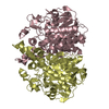
| Title | Crystal structure of beta-ketoacyl synthase from brucella melitensis with FOL 0758, (1-methyl-1h-indazol-3-yl) methanol | ||||||
 Components Components | Beta-ketoacyl synthase | ||||||
 Keywords Keywords |  TRANSFERASE / SSGCID / ALS COLLABORATIVE CRYSTALLOGRAPHY / BETA-KETOACYL SYNTHASE / BRUCELLA MELITENSIS / FRAGMENTS OF LIFE / Structural Genomics / Seattle Structural Genomics Center for Infectious Disease TRANSFERASE / SSGCID / ALS COLLABORATIVE CRYSTALLOGRAPHY / BETA-KETOACYL SYNTHASE / BRUCELLA MELITENSIS / FRAGMENTS OF LIFE / Structural Genomics / Seattle Structural Genomics Center for Infectious Disease | ||||||
| Function / homology |  Function and homology information Function and homology informationbeta-ketoacyl-[acyl-carrier-protein] synthase I / 3-oxoacyl-[acyl-carrier-protein] synthase activity / fatty acid biosynthetic process Similarity search - Function | ||||||
| Biological species |  Brucella melitensis biovar Abortus (bacteria) Brucella melitensis biovar Abortus (bacteria) | ||||||
| Method | X-RAY DIFFRACTION / SYNCHROTRON / molecular replacement / Resolution: 1.25 Å | ||||||
 Authors Authors | Seattle Structural Genomics Center for Infectious Disease (SSGCID) | ||||||
 Citation Citation | Journal: Proteins / Year: 2020 Title: Structural characterization of beta-ketoacyl ACP synthase I bound to platencin and fragment screening molecules at two substrate binding sites. Authors: Patterson, E.I. / Nanson, J.D. / Abendroth, J. / Bryan, C. / Sankaran, B. / Myler, P.J. / Forwood, J.K. | ||||||
| History |
|
- Structure visualization
| Structure viewer | Molecule: MolmilJmol/JSmol |
|---|
- Downloads & links
-Download
| PDBx/mmCIF format | 3mqd.cif.gz | 188.7 KB | Display | PDBx/mmCIF format |
|---|---|---|---|---|
| PDB format | pdb3mqd.ent.gz | 146.6 KB | Display | PDB format |
| PDBx/mmJSON format | 3mqd.json.gz | Tree view | PDBx/mmJSON format | |
| Others |  Other downloads Other downloads |
-Validation report
| Arichive directory | https://data.pdbj.org/pub/pdb/validation_reports/mq/3mqdftp://data.pdbj.org/pub/pdb/validation_reports/mq/3mqd | HTTPS FTP |
|---|
-Related structure data
| Related structure data | 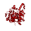 3lrfSC 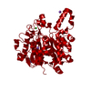 3u0eC 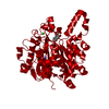 3u0fC  4jv3C S: Starting model for refinement C: citing same article ( |
|---|---|
| Similar structure data | |
| Other databases |


-Links
 PDBj
PDBj

- Assembly
| Deposited unit | 
| ||||||||
|---|---|---|---|---|---|---|---|---|---|
| 1 | 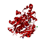
| ||||||||
| Unit cell |
| ||||||||
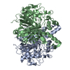
| Details | BIOMOLECULE: NULL SEE REMARK 350 FOR THE AUTHOR PROVIDED AND/OR PROGRAM GENERATED ASSEMBLY INFORMATION FOR THE STRUCTURE IN THIS ENTRY. THE REMARK MAY ALSO PROVIDE INFORMATION ON BURIED SURFACE AREA. REMARK: BIOLOGICAL UNIT IS A DIMER |
-Components
| #1: Protein | Mass: 45643.273 Da / Num. of mol.: 1 Source method: isolated from a genetically manipulated source Source: (gene. exp.) Brucella melitensis biovar Abortus (bacteria)Strain: ABORTUS 2308 / Gene: fabB, BAB1_2173 / Plasmid: AVA0421 / Production host: Escherichia coli (E. coli) / Strain (production host): BL21(DE3)References: UniProt: Q2YQQ9, glucosamine-phosphate N-acetyltransferase | ||||||
|---|---|---|---|---|---|---|---|
| #2: Chemical |   Mass: 22.990 Da / Num. of mol.: 2 / Source method: obtained synthetically / Formula: Na Mass: 22.990 Da / Num. of mol.: 2 / Source method: obtained synthetically / Formula: Na#3: Chemical | ChemComp-CL / | Chloride  Mass: 35.453 Da / Num. of mol.: 1 / Source method: obtained synthetically / Formula: Cl Mass: 35.453 Da / Num. of mol.: 1 / Source method: obtained synthetically / Formula: Cl#4: Chemical | ChemComp-3MQ / ( |   Mass: 181.212 Da / Num. of mol.: 1 / Source method: obtained synthetically / Formula: C8H7NO2S Mass: 181.212 Da / Num. of mol.: 1 / Source method: obtained synthetically / Formula: C8H7NO2S#5: Water | ChemComp-HOH / | Water Mass: 18.015 Da / Num. of mol.: 454 / Source method: isolated from a natural source / Formula: H2O Mass: 18.015 Da / Num. of mol.: 454 / Source method: isolated from a natural source / Formula: H2O |
-Experimental details
-Experiment
| Experiment | Method: X-RAY DIFFRACTION / Number of used crystals: 1 |
|---|
- Sample preparation
| Crystal | Density Matthews: 2.25 Å3/Da / Density % sol: 45.29 % |
|---|---|
| Crystal grow | Temperature: 290 K / pH: 8.5 Details: MD PACT SCREEN, H12: 20% PEG 3350, 100MM BISTRISPROPANE PH 8.5, 200MM NA-MALONATE; CRYSTAL SOAKED 100MM MES PH 6.5, 250MM NACL, 30% PEG 3350, 10% GLYCEROL; BRABA.00113.A AT 22.9MG/ML, VAPOR ...Details: MD PACT SCREEN, H12: 20% PEG 3350, 100MM BISTRISPROPANE PH 8.5, 200MM NA-MALONATE; CRYSTAL SOAKED 100MM MES PH 6.5, 250MM NACL, 30% PEG 3350, 10% GLYCEROL; BRABA.00113.A AT 22.9MG/ML, VAPOR DIFFUSION, SITTING DROP, TEMPERATURE 290K |
-Data collection
| Diffraction | Mean temperature: 100 K |
|---|---|
| Diffraction source | Source: SYNCHROTRON / Site: ALS  / Beamline: 5.0.1 / Wavelength: 0.9774 / Beamline: 5.0.1 / Wavelength: 0.9774 |
| Detector | Type: ADSC QUANTUM Q315R / Detector: CCD / Date: Apr 2, 2010 |
| Radiation | Protocol: SINGLE WAVELENGTH / Scattering type: x-ray |
| Radiation wavelength | Wavelength: 0.9774 Å / Relative weight: 1 |
| Reflection | Resolution: 1.25→4181 Å / Num. obs: 111226 / % possible obs: 99.7 % / Observed criterion σ(I): -3 / Biso Wilson estimate: 13.34 Å2 / Rmerge(I) obs: 0.091 / Net I/σ(I): 12.07 |
| Reflection shell | Resolution: 1.25→1.28 Å / Rmerge(I) obs: 0.404 / Mean I/σ(I) obs: 4 / % possible all: 99.1 |
- Processing
| Software |
| ||||||||||||||||||||||||||||||||||||||||||||||||||||||||||||||||||||||||||||||||||||||||||||||||||||||||||||||||||||||||||||||||||||||||||||||||||||||||||||||||||||||||||
|---|---|---|---|---|---|---|---|---|---|---|---|---|---|---|---|---|---|---|---|---|---|---|---|---|---|---|---|---|---|---|---|---|---|---|---|---|---|---|---|---|---|---|---|---|---|---|---|---|---|---|---|---|---|---|---|---|---|---|---|---|---|---|---|---|---|---|---|---|---|---|---|---|---|---|---|---|---|---|---|---|---|---|---|---|---|---|---|---|---|---|---|---|---|---|---|---|---|---|---|---|---|---|---|---|---|---|---|---|---|---|---|---|---|---|---|---|---|---|---|---|---|---|---|---|---|---|---|---|---|---|---|---|---|---|---|---|---|---|---|---|---|---|---|---|---|---|---|---|---|---|---|---|---|---|---|---|---|---|---|---|---|---|---|---|---|---|---|---|---|---|---|
| Refinement | Method to determine structure: molecular replacement Starting model: native structure, 3LRF Resolution: 1.25→41.81 Å / Cor.coef. Fo:Fc: 0.979 / Cor.coef. Fo:Fc free: 0.973 / SU B: 0.821 / SU ML: 0.017 Isotropic thermal model: anisotropic, some solvent atoms left isotropic Cross valid method: THROUGHOUT / σ(F): 0 / ESU R: 0.032 / ESU R Free: 0.031 / Stereochemistry target values: MAXIMUM LIKELIHOOD / Details: HYDROGENS HAVE BEEN ADDED IN THE
| ||||||||||||||||||||||||||||||||||||||||||||||||||||||||||||||||||||||||||||||||||||||||||||||||||||||||||||||||||||||||||||||||||||||||||||||||||||||||||||||||||||||||||
| Solvent computation | Ion probe radii: 0.8 Å / Shrinkage radii: 0.8 Å / VDW probe radii: 1.4 Å / Solvent model: MASK | ||||||||||||||||||||||||||||||||||||||||||||||||||||||||||||||||||||||||||||||||||||||||||||||||||||||||||||||||||||||||||||||||||||||||||||||||||||||||||||||||||||||||||
| Displacement parameters | Biso mean: 7.42 Å2
| ||||||||||||||||||||||||||||||||||||||||||||||||||||||||||||||||||||||||||||||||||||||||||||||||||||||||||||||||||||||||||||||||||||||||||||||||||||||||||||||||||||||||||
| Refinement step | Cycle: LAST / Resolution: 1.25→41.81 Å
| ||||||||||||||||||||||||||||||||||||||||||||||||||||||||||||||||||||||||||||||||||||||||||||||||||||||||||||||||||||||||||||||||||||||||||||||||||||||||||||||||||||||||||
| Refine LS restraints |
| ||||||||||||||||||||||||||||||||||||||||||||||||||||||||||||||||||||||||||||||||||||||||||||||||||||||||||||||||||||||||||||||||||||||||||||||||||||||||||||||||||||||||||
| LS refinement shell | Resolution: 1.25→1.28 Å / Total num. of bins used: 20
|
