 Movie
Movie Controller


+ Open data

- Basic information
| Entry | Database: PDB / ID: 3mpx | ||||||
|---|---|---|---|---|---|---|---|
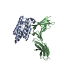
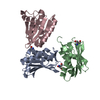
| Title | Crystal structure of the DH and PH-1 domains of human FGD5 | ||||||
 Components Components | FYVE, RhoGEF and PH domain-containing protein 5 | ||||||
 Keywords Keywords | LIPID BINDING PROTEIN /  Structural Genomics Consortium / DH domain / PH domain / SGC Structural Genomics Consortium / DH domain / PH domain / SGC | ||||||
| Function / homology |  Function and homology information Function and homology informationguanyl-nucleotide exchange factor activity => GO:0005085 / filopodium assembly / regulation of GTPase activity / ruffle / cytoskeleton organization / guanyl-nucleotide exchange factor activity / small GTPase binding / ruffle membrane / lamellipodium / regulation of cell shape ...guanyl-nucleotide exchange factor activity => GO:0005085 / filopodium assembly / regulation of GTPase activity / ruffle / cytoskeleton organization / guanyl-nucleotide exchange factor activity / small GTPase binding / ruffle membrane / lamellipodium / regulation of cell shape / actin cytoskeleton organization / early endosome / cytoskeleton / Golgi apparatus / endoplasmic reticulum / metal ion binding / plasma membrane / cytoplasmSimilarity search - Function | ||||||
| Biological species |  Homo sapiens (human) Homo sapiens (human) | ||||||
| Method | X-RAY DIFFRACTION / SYNCHROTRON / MAD / mad / Resolution: 2.8 Å | ||||||
 Authors Authors | Shen, Y. / Nedyalkova, L. / Tong, Y. / Tempel, W. / Crombet, L. / Arrowsmith, C.H. / Edwards, A.M. / Bountra, C. / Weigelt, J. / Bochkarev, A. ...Shen, Y. / Nedyalkova, L. / Tong, Y. / Tempel, W. / Crombet, L. / Arrowsmith, C.H. / Edwards, A.M. / Bountra, C. / Weigelt, J. / Bochkarev, A. / Park, H. / Structural Genomics Consortium (SGC) | ||||||
 Citation Citation | Journal: TO BE PUBLISHED Title: Crystal structure of the DH and PH-1 domains of human FGD5 Authors: Shen, Y. / Nedyalkova, L. / Tong, Y. / Tempel, W. / Crombet, L. / Arrowsmith, C.H. / Edwards, A.M. / Bountra, C. / Weigelt, J. / Bochkarev, A. / Park, H. | ||||||
| History |
|
- Structure visualization
| Structure viewer | Molecule: MolmilJmol/JSmol |
|---|
- Downloads & links
-Download
| PDBx/mmCIF format | 3mpx.cif.gz | 72 KB | Display | PDBx/mmCIF format |
|---|---|---|---|---|
| PDB format | pdb3mpx.ent.gz | 54 KB | Display | PDB format |
| PDBx/mmJSON format | 3mpx.json.gz | Tree view | PDBx/mmJSON format | |
| Others |  Other downloads Other downloads |
-Validation report
| Arichive directory | https://data.pdbj.org/pub/pdb/validation_reports/mp/3mpxftp://data.pdbj.org/pub/pdb/validation_reports/mp/3mpx | HTTPS FTP |
|---|
-Related structure data
| Similar structure data |
|---|


-Links
 PDBj
PDBj

- Assembly
| Deposited unit | 
| ||||||||
|---|---|---|---|---|---|---|---|---|---|
| 1 |
| ||||||||
| Unit cell |
|
-Components
| #1: Protein | Mass: 50303.230 Da / Num. of mol.: 1 / Fragment: UNP Residues 889-1304 Source method: isolated from a genetically manipulated source Source: (gene. exp.) Homo sapiens (human) / Gene: FGD5, ZFYVE23 / Plasmid: pET28-MHL / Production host:  Escherichia coli (E. coli) / Strain (production host): BL21(DE3)-V2R-pRARE2 / References: UniProt: Q6ZNL6 Escherichia coli (E. coli) / Strain (production host): BL21(DE3)-V2R-pRARE2 / References: UniProt: Q6ZNL6 | ||
|---|---|---|---|
| #2: Chemical | ChemComp-UNX /   Num. of mol.: 11 / Source method: obtained synthetically Num. of mol.: 11 / Source method: obtained syntheticallySequence details | THE PROTEIN USED FOR CRYSTALLIZATION CONTAINED AMINO-ACIDS AS GIVEN IN DBREF. THIS PROTEIN LIKELY ...THE PROTEIN USED FOR CRYSTALLIZ | |
-Experimental details
-Experiment
| Experiment | Method: X-RAY DIFFRACTION / Number of used crystals: 1 |
|---|
- Sample preparation
| Crystal | Density Matthews: 2.4 Å3/Da / Density % sol: 48.6 % |
|---|---|
| Crystal grow | Temperature: 291 K / Method: vapor diffusion, sitting drop / pH: 7.5 Details: 25% w/v PEG 3350, 0.2M lithium sulfate, 0.1M HEPES pH 7.5. 1:100 trypsin was also added. vapor diffusion, sitting drop, temperature 291K, VAPOR DIFFUSION, SITTING DROP |
-Data collection
| Diffraction | Mean temperature: 100 K | ||||||||||||||||||||||||||||||||||||||||||||||||||||||||||||||||||||||||||||||||||||||||||||||||||||||||||||||||||||||||||||||
|---|---|---|---|---|---|---|---|---|---|---|---|---|---|---|---|---|---|---|---|---|---|---|---|---|---|---|---|---|---|---|---|---|---|---|---|---|---|---|---|---|---|---|---|---|---|---|---|---|---|---|---|---|---|---|---|---|---|---|---|---|---|---|---|---|---|---|---|---|---|---|---|---|---|---|---|---|---|---|---|---|---|---|---|---|---|---|---|---|---|---|---|---|---|---|---|---|---|---|---|---|---|---|---|---|---|---|---|---|---|---|---|---|---|---|---|---|---|---|---|---|---|---|---|---|---|---|---|
| Diffraction source | Source: SYNCHROTRON / Site: APS  / Beamline: 19-ID / Wavelength: 0.97942, 0.97934 / Beamline: 19-ID / Wavelength: 0.97942, 0.97934 | ||||||||||||||||||||||||||||||||||||||||||||||||||||||||||||||||||||||||||||||||||||||||||||||||||||||||||||||||||||||||||||||
| Detector | Type: ADSC QUANTUM 315r / Detector: CCD / Date: Mar 25, 2010 | ||||||||||||||||||||||||||||||||||||||||||||||||||||||||||||||||||||||||||||||||||||||||||||||||||||||||||||||||||||||||||||||
| Radiation | Protocol: MAD / Monochromatic (M) / Laue (L): M / Scattering type: x-ray | ||||||||||||||||||||||||||||||||||||||||||||||||||||||||||||||||||||||||||||||||||||||||||||||||||||||||||||||||||||||||||||||
| Radiation wavelength |
| ||||||||||||||||||||||||||||||||||||||||||||||||||||||||||||||||||||||||||||||||||||||||||||||||||||||||||||||||||||||||||||||
| Reflection | Resolution: 2.8→30 Å / Num. obs: 9116 / % possible obs: 99.8 % / Redundancy: 6.9 % / Rmerge(I) obs: 0.142 / Χ2: 1.237 / Net I/σ(I): 5.5 | ||||||||||||||||||||||||||||||||||||||||||||||||||||||||||||||||||||||||||||||||||||||||||||||||||||||||||||||||||||||||||||||
| Reflection shell |
|
-Phasing
| Phasing | Method: mad |
|---|
- Processing
| Software |
| ||||||||||||||||||||||||||||||||||||||||||||||||||||||||||||||||||||||||||||||||||||||||||||||||||||||||||||||||||
|---|---|---|---|---|---|---|---|---|---|---|---|---|---|---|---|---|---|---|---|---|---|---|---|---|---|---|---|---|---|---|---|---|---|---|---|---|---|---|---|---|---|---|---|---|---|---|---|---|---|---|---|---|---|---|---|---|---|---|---|---|---|---|---|---|---|---|---|---|---|---|---|---|---|---|---|---|---|---|---|---|---|---|---|---|---|---|---|---|---|---|---|---|---|---|---|---|---|---|---|---|---|---|---|---|---|---|---|---|---|---|---|---|---|---|---|
| Refinement | Method to determine structure: MAD / Resolution: 2.8→29.71 Å / Cor.coef. Fo:Fc: 0.894 / Cor.coef. Fo:Fc free: 0.842 / Cross valid method: THROUGHOUT / σ(F): 0 Details: Model was refined against inflection wavelength data
| ||||||||||||||||||||||||||||||||||||||||||||||||||||||||||||||||||||||||||||||||||||||||||||||||||||||||||||||||||
| Displacement parameters | Biso mean: 29.79 Å2
| ||||||||||||||||||||||||||||||||||||||||||||||||||||||||||||||||||||||||||||||||||||||||||||||||||||||||||||||||||
| Refine analyze | Luzzati coordinate error obs: 0.376 Å | ||||||||||||||||||||||||||||||||||||||||||||||||||||||||||||||||||||||||||||||||||||||||||||||||||||||||||||||||||
| Refinement step | Cycle: LAST / Resolution: 2.8→29.71 Å
| ||||||||||||||||||||||||||||||||||||||||||||||||||||||||||||||||||||||||||||||||||||||||||||||||||||||||||||||||||
| Refine LS restraints |
| ||||||||||||||||||||||||||||||||||||||||||||||||||||||||||||||||||||||||||||||||||||||||||||||||||||||||||||||||||
| LS refinement shell | Resolution: 2.8→3.13 Å / Total num. of bins used: 5
|
