 Movie
Movie Controller

[English] 日本語
 Yorodumi
- PDB-3mlh: Crystal structure of the 2009 H1N1 influenza virus hemagglutinin ... -
+ Open data

- Basic information
| Entry | Database: PDB / ID: 3mlh | ||||||
|---|---|---|---|---|---|---|---|
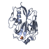
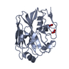
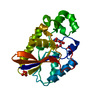
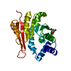
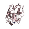
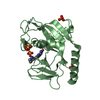
| Title | Crystal structure of the 2009 H1N1 influenza virus hemagglutinin receptor-binding domain | ||||||
 Components Components | Hemagglutinin | ||||||
 Keywords Keywords | VIRAL PROTEIN / hemagglutinin / receptor-binding domain / lectin / antigen | ||||||
| Function / homology |  Function and homology informationviral budding from plasma membrane / clathrin-dependent endocytosis of virus by host cell / membrane => GO:0016020 / host cell surface receptor binding / apical plasma membrane / fusion of virus membrane with host plasma membrane / fusion of virus membrane with host endosome membrane / viral envelope / virion attachment to host cell / host cell plasma membrane / virion membrane Function and homology informationviral budding from plasma membrane / clathrin-dependent endocytosis of virus by host cell / membrane => GO:0016020 / host cell surface receptor binding / apical plasma membrane / fusion of virus membrane with host plasma membrane / fusion of virus membrane with host endosome membrane / viral envelope / virion attachment to host cell / host cell plasma membrane / virion membraneSimilarity search - Function | ||||||
| Biological species |   Influenza A virus Influenza A virus | ||||||
| Method | X-RAY DIFFRACTION / SYNCHROTRON / MOLECULAR REPLACEMENT / molecular replacement / Resolution: 2.09 Å | ||||||
 Authors Authors | DuBois, R.M. / Aguilar-Yanez, J.M. / Mendoza-Ochoa, G.I. / Schultz-Cherry, S. / Alvarez, M.M. / White, S.W. / Russell, C.J. | ||||||
 Citation Citation | Journal: J.Virol. / Year: 2011 Title: The Receptor-Binding Domain of Influenza Virus Hemagglutinin Produced in Escherichia coli Folds into Its Native, Immunogenic Structure. Authors: Dubois, R.M. / Aguilar-Yanez, J.M. / Mendoza-Ochoa, G.I. / Oropeza-Almazan, Y. / Schultz-Cherry, S. / Alvarez, M.M. / White, S.W. / Russell, C.J. | ||||||
| History |
|
- Structure visualization
| Structure viewer | Molecule: MolmilJmol/JSmol |
|---|
- Downloads & links
-Download
| PDBx/mmCIF format | 3mlh.cif.gz | 178.1 KB | Display | PDBx/mmCIF format |
|---|---|---|---|---|
| PDB format | pdb3mlh.ent.gz | 141.8 KB | Display | PDB format |
| PDBx/mmJSON format | 3mlh.json.gz | Tree view | PDBx/mmJSON format | |
| Others |  Other downloads Other downloads |
-Validation report
| Arichive directory | https://data.pdbj.org/pub/pdb/validation_reports/ml/3mlhftp://data.pdbj.org/pub/pdb/validation_reports/ml/3mlh | HTTPS FTP |
|---|
-Related structure data
| Related structure data | 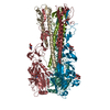 1ruyS S: Starting model for refinement |
|---|---|
| Similar structure data |


-Links
 PDBj
PDBj


- Assembly
| Deposited unit | 
| ||||||||
|---|---|---|---|---|---|---|---|---|---|
| 1 | 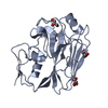
| ||||||||
| 2 | 
| ||||||||
| Unit cell |
|
-Components
| #1: Protein | Mass: 26414.424 Da / Num. of mol.: 2 / Fragment: receptor-binding domain (UNP residues 63-286) Source method: isolated from a genetically manipulated source Source: (gene. exp.) Influenza A virus / Strain: A/Mexico/4603/2009(H1N1) / Gene: HA / Plasmid: pJexpress404 / Production host:  Escherichia coli (E. coli) / Strain (production host): C41 strain / References: UniProt: C4RSQ3 Escherichia coli (E. coli) / Strain (production host): C41 strain / References: UniProt: C4RSQ3#2: Chemical | ChemComp-GOL / Glycerol  Mass: 92.094 Da / Num. of mol.: 6 / Source method: obtained synthetically / Formula: C3H8O3 Mass: 92.094 Da / Num. of mol.: 6 / Source method: obtained synthetically / Formula: C3H8O3#3: Water | ChemComp-HOH / | Water Mass: 18.015 Da / Num. of mol.: 193 / Source method: isolated from a natural source / Formula: H2O Mass: 18.015 Da / Num. of mol.: 193 / Source method: isolated from a natural source / Formula: H2O |
|---|
-Experimental details
-Experiment
| Experiment | Method: X-RAY DIFFRACTION / Number of used crystals: 1 |
|---|
- Sample preparation
| Crystal | Density Matthews: 2.09 Å3/Da / Density % sol: 41.24 % |
|---|---|
| Crystal grow | Temperature: 291 K / Method: vapor diffusion, hanging drop / pH: 8.5 Details: PEG 2000MME, Tris pH 8.5, VAPOR DIFFUSION, HANGING DROP, temperature 291K |
-Data collection
| Diffraction | Mean temperature: 100 K |
|---|---|
| Diffraction source | Source: SYNCHROTRON / Site: ALS  / Beamline: 8.2.1 / Wavelength: 1 Å / Beamline: 8.2.1 / Wavelength: 1 Å |
| Detector | Type: ADSC QUANTUM 315r / Detector: CCD / Date: Mar 4, 2010 |
| Radiation | Protocol: SINGLE WAVELENGTH / Monochromatic (M) / Laue (L): M / Scattering type: x-ray |
| Radiation wavelength | Wavelength: 1 Å / Relative weight: 1 |
| Reflection | Resolution: 2.1→40 Å / Num. all: 25593 / Num. obs: 25588 / % possible obs: 100 % / Observed criterion σ(F): 0 / Observed criterion σ(I): 12.73 / Redundancy: 5 % / Biso Wilson estimate: 19.628 Å2 / Rmerge(I) obs: 0.127 / Χ2: 1.147 / Net I/σ(I): 8.3 |
| Reflection shell | Resolution: 2.1→2.18 Å / Redundancy: 3.9 % / Rmerge(I) obs: 0.373 / Mean I/σ(I) obs: 3.83 / Num. unique all: 2518 / Χ2: 1.123 / % possible all: 100 |
-Phasing
| Phasing | Method: molecular replacement | |||||||||
|---|---|---|---|---|---|---|---|---|---|---|
| Phasing MR | Rfactor: 44.07 / Model details: Phaser MODE: MR_AUTO
|
- Processing
| Software |
| |||||||||||||||||||||||||||||||||||||||||||||||||||||||||||||||||||||||||||
|---|---|---|---|---|---|---|---|---|---|---|---|---|---|---|---|---|---|---|---|---|---|---|---|---|---|---|---|---|---|---|---|---|---|---|---|---|---|---|---|---|---|---|---|---|---|---|---|---|---|---|---|---|---|---|---|---|---|---|---|---|---|---|---|---|---|---|---|---|---|---|---|---|---|---|---|---|
| Refinement | Method to determine structure: MOLECULAR REPLACEMENT Starting model: PDB entry 1RUY Resolution: 2.09→39.72 Å / Cor.coef. Fo:Fc: 0.947 / Cor.coef. Fo:Fc free: 0.925 / Occupancy max: 1 / Occupancy min: 0.5 / SU B: 9.233 / SU ML: 0.113 / Cross valid method: THROUGHOUT / σ(F): 0 / ESU R: 0.229 / ESU R Free: 0.169 / Stereochemistry target values: MAXIMUM LIKELIHOOD Details: HYDROGENS HAVE BEEN ADDED IN THE RIDING POSITIONS. U VALUES: RESIDUAL ONLY
| |||||||||||||||||||||||||||||||||||||||||||||||||||||||||||||||||||||||||||
| Solvent computation | Ion probe radii: 0.8 Å / Shrinkage radii: 0.8 Å / VDW probe radii: 1.4 Å / Solvent model: MASK | |||||||||||||||||||||||||||||||||||||||||||||||||||||||||||||||||||||||||||
| Displacement parameters | Biso max: 43.65 Å2 / Biso mean: 19.628 Å2 / Biso min: 5.58 Å2
| |||||||||||||||||||||||||||||||||||||||||||||||||||||||||||||||||||||||||||
| Refinement step | Cycle: LAST / Resolution: 2.09→39.72 Å
| |||||||||||||||||||||||||||||||||||||||||||||||||||||||||||||||||||||||||||
| Refine LS restraints |
| |||||||||||||||||||||||||||||||||||||||||||||||||||||||||||||||||||||||||||
| LS refinement shell | Resolution: 2.09→2.144 Å / Total num. of bins used: 20
| |||||||||||||||||||||||||||||||||||||||||||||||||||||||||||||||||||||||||||
| Refinement TLS params. | Method: refined / Refine-ID: X-RAY DIFFRACTION
| |||||||||||||||||||||||||||||||||||||||||||||||||||||||||||||||||||||||||||
| Refinement TLS group |
|
