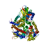
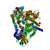
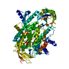
Entry Database : PDB / ID : 3ml9Title Discovery of the Highly Potent PI3K/mTOR Dual Inhibitor PF-04691502 through Structure Based Drug Design Phosphatidylinositol-4,5-bisphosphate 3-kinase catalytic subunit gamma isoform Keywords / / / Function / homology Function Domain/homology Component
/ / / / / / / / / / / / / / / / / / / / / / / / / / / / / / / / / / / / / / / / / / / / / / / / / / / / / / / / / / / / / / / / / / / / / / / / / / / / / / / / / / / / / / / / / / / / / / / / / / / / / / / / / / / / / / / / / / / / / / / / / / / / / / / Biological species Homo sapiens (human)Method / / / Resolution : 2.55 Å Authors Knighton, D.R. Journal : ACS Med Chem Lett / Year : 2013Title : Discovery of the Highly Potent PI3K/mTOR Dual Inhibitor PF-04979064 through Structure-Based Drug Design.Authors: Cheng, H. / Li, C. / Bailey, S. / Baxi, S.M. / Goulet, L. / Guo, L. / Hoffman, J. / Jiang, Y. / Johnson, T.O. / Johnson, T.W. / Knighton, D.R. / Li, J. / Liu, K.K. / Liu, Z. / Marx, M.A. / ... Authors : Cheng, H. / Li, C. / Bailey, S. / Baxi, S.M. / Goulet, L. / Guo, L. / Hoffman, J. / Jiang, Y. / Johnson, T.O. / Johnson, T.W. / Knighton, D.R. / Li, J. / Liu, K.K. / Liu, Z. / Marx, M.A. / Walls, M. / Wells, P.A. / Yin, M.J. / Zhu, J. / Zientek, M. History Deposition Apr 16, 2010 Deposition site / Processing site Revision 1.0 Jun 2, 2010 Provider / Type Revision 1.1 Jul 13, 2011 Group Revision 1.2 Jul 9, 2014 Group Revision 1.3 Feb 21, 2024 Group / Database references / Derived calculationsCategory chem_comp_atom / chem_comp_bond ... chem_comp_atom / chem_comp_bond / database_2 / struct_ref_seq_dif / struct_site Item _database_2.pdbx_DOI / _database_2.pdbx_database_accession ... _database_2.pdbx_DOI / _database_2.pdbx_database_accession / _struct_ref_seq_dif.details / _struct_site.pdbx_auth_asym_id / _struct_site.pdbx_auth_comp_id / _struct_site.pdbx_auth_seq_id
Show all Show less
 Movie
Movie Controller

 Yorodumi
 Open data
 Basic information
 Components
 Keywords
 inhibition / TRANSFERASE-TRANSFERASE INHIBITOR complex
 Function and homology information
 Authors
 Citation
 Structure visualization
 Downloads & links
 Other downloads
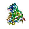

 PDBj
PDBj


 Assembly


 Mass: 425.481 Da / Num. of mol.: 1 / Source method: obtained synthetically / Formula: C22H27N5O4
 Mass: 18.015 Da / Num. of mol.: 1 / Source method: isolated from a natural source / Formula: H2O
 Sample preparation
 / Beamline: 5.0.2 / Wavelength: 1 Å
 Processing